Baars Matthijs J D, Floor Evelien, Sinha Neeraj, Ter Linde José J M, van Dam Stephanie, Amini Mojtaba, Nijman Isaäc J, Ten Hove Joren R, Drylewicz Julia, Offerhaus G Johan A, Laclé Miangela M, Oldenburg Bas, Vercoulen Yvonne
Center for Molecular Medicine, University Medical Center Utrecht, Utrecht University, Universiteitsweg 100, CX, Utrecht 3584, the Netherlands.
Department of Gastroenterology and Hepatology, University Medical Center Utrecht, Utrecht University, Heidelberglaan 100, CX, Utrecht 3584, the Netherlands.
iScience. 2024 Jul 20;27(8):110550. doi: 10.1016/j.isci.2024.110550. eCollection 2024 Aug 16.
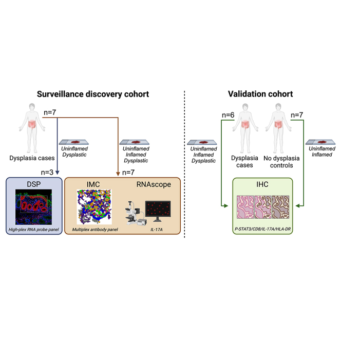
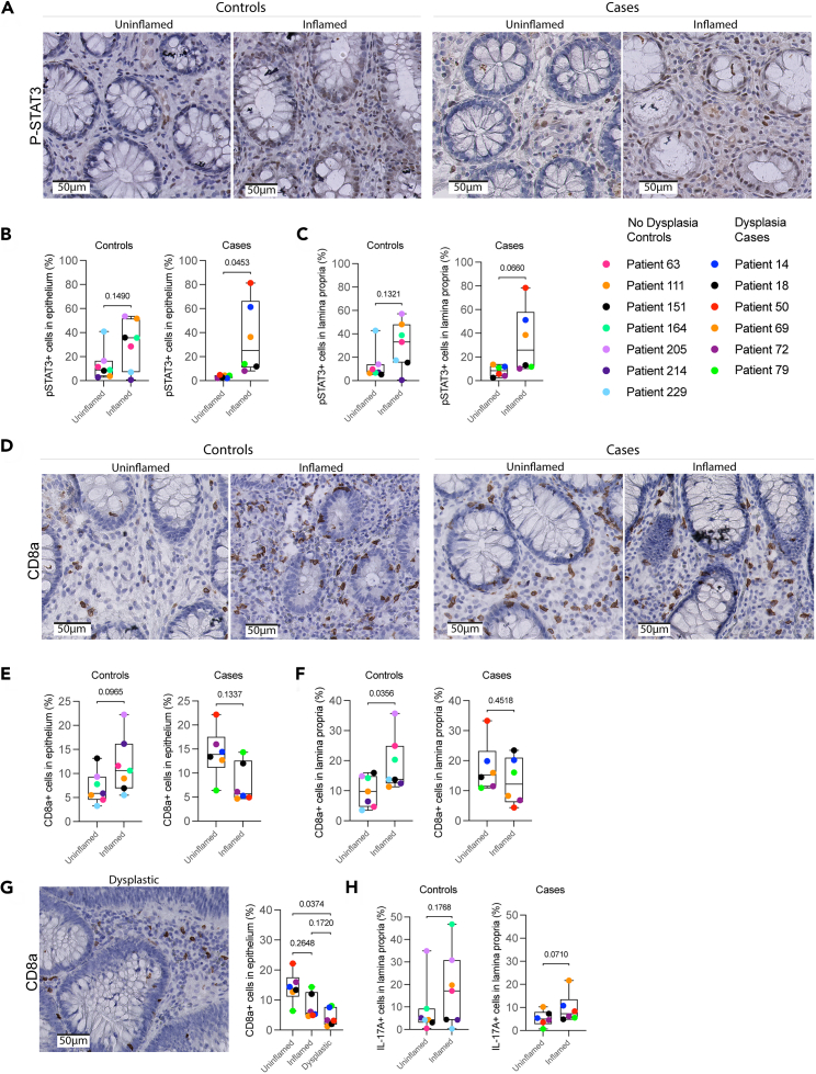
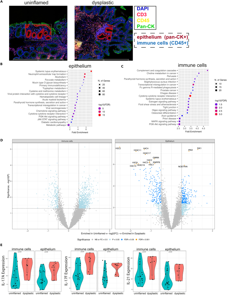
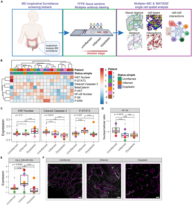
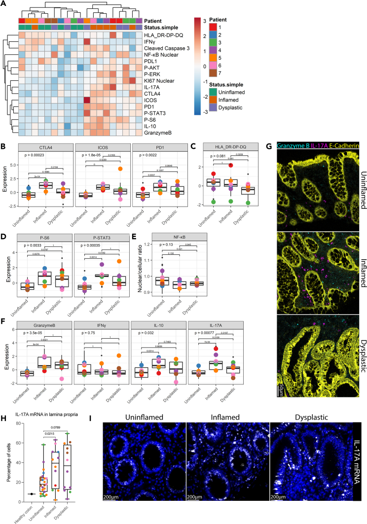
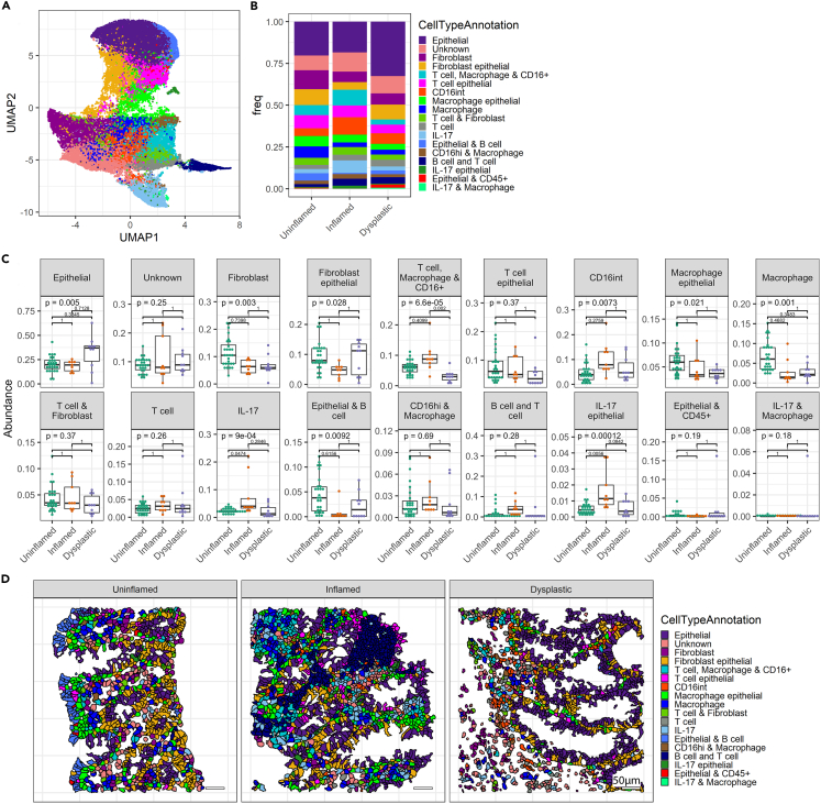
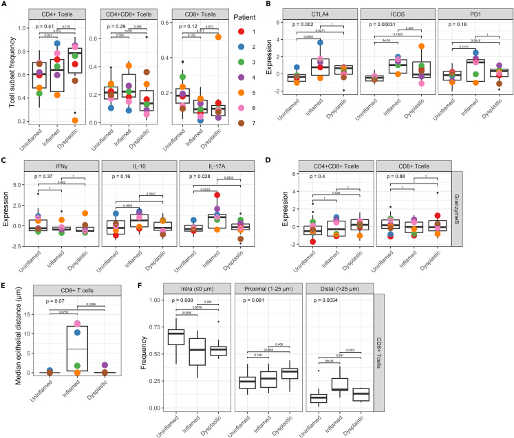
Patients with long-standing inflammatory bowel disease (IBD) face an increased risk of developing colitis-associated cancer (CAC). Although IBD-induced prolonged inflammation seems to be involved in CAC pathogenesis, the specific molecular changes that contribute remain unknown. Here, we applied digital spatial RNA profiling, RNAscope, and imaging mass cytometry to examine paired uninflamed, inflamed, and early dysplastic mucosa of patients with IBD. We observed robust type 3 (IL-17) responses during inflammation, accompanied by elevated JAK-STAT signaling and phosphorylated STAT3 (P-STAT3) levels, with both inflamed and dysplastic mucosa displaying immune cell activation. Higher stromal P-STAT3 was detected in uninflamed and inflamed mucosa of patients who eventually developed dysplasia. CD8a T cells did not infiltrate inflamed or dysplastic epithelial regions in these patients, while control patients showed elevated CD8a in inflamed mucosa. Our study reveals distinct inflammatory patterns throughout CAC development, marked by an activated IL-17 pathway, engaged STAT3, and diminished cytotoxic T cell infiltration.
患有长期炎症性肠病(IBD)的患者患结肠炎相关癌症(CAC)的风险增加。虽然IBD诱导的长期炎症似乎参与了CAC的发病机制,但具体的分子变化仍不清楚。在这里,我们应用数字空间RNA分析、RNAscope和成像质谱流式细胞术来检查IBD患者的配对未发炎、发炎和早期发育异常的黏膜。我们观察到炎症期间强大的3型(IL-17)反应,伴随着JAK-STAT信号通路的升高和磷酸化STAT3(P-STAT3)水平的升高,发炎和发育异常的黏膜均显示免疫细胞活化。在最终发生发育异常的患者的未发炎和发炎黏膜中检测到较高的基质P-STAT3。在这些患者中,CD8a T细胞未浸润发炎或发育异常的上皮区域,而对照患者发炎黏膜中的CD8a升高。我们的研究揭示了整个CAC发展过程中不同的炎症模式,其特征是IL-17通路激活、STAT3参与以及细胞毒性T细胞浸润减少。