Molecular Cancer Research, Center for Molecular Medicine, University Medical Center Utrecht, Utrecht University, 3584, CX, Utrecht, The Netherlands.
Oncode Institute, Utrecht, The Netherlands.
BMC Biol. 2021 May 11;19(1):99. doi: 10.1186/s12915-021-01043-y.
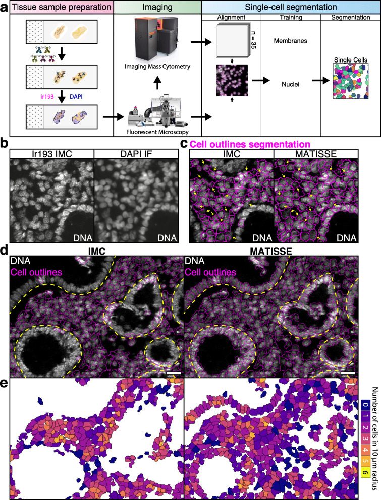
Visualizing and quantifying cellular heterogeneity is of central importance to study tissue complexity, development, and physiology and has a vital role in understanding pathologies. Mass spectrometry-based methods including imaging mass cytometry (IMC) have in recent years emerged as powerful approaches for assessing cellular heterogeneity in tissues. IMC is an innovative multiplex imaging method that combines imaging using up to 40 metal conjugated antibodies and provides distributions of protein markers in tissues with a resolution of 1 μm area. However, resolving the output signals of individual cells within the tissue sample, i.e., single cell segmentation, remains challenging. To address this problem, we developed MATISSE (iMaging mAss cyTometry mIcroscopy Single cell SegmEntation), a method that combines high-resolution fluorescence microscopy with the multiplex capability of IMC into a single workflow to achieve improved segmentation over the current state-of-the-art.
MATISSE results in improved quality and quantity of segmented cells when compared to IMC-only segmentation in sections of heterogeneous tissues. Additionally, MATISSE enables more complete and accurate identification of epithelial cells, fibroblasts, and infiltrating immune cells in densely packed cellular areas in tissue sections. MATISSE has been designed based on commonly used open-access tools and regular fluorescence microscopy, allowing easy implementation by labs using multiplex IMC into their analysis methods.
MATISSE allows segmentation of densely packed cellular areas and provides a qualitative and quantitative improvement when compared to IMC-based segmentation. We expect that implementing MATISSE into tissue section analysis pipelines will yield improved cell segmentation and enable more accurate analysis of the tissue microenvironment in epithelial tissue pathologies, such as autoimmunity and cancer.
可视化和量化细胞异质性对于研究组织复杂性、发育和生理学至关重要,对于理解病理学也具有重要作用。基于质谱的方法,包括成像质谱流式细胞术(IMC),近年来已成为评估组织中细胞异质性的有力方法。IMC 是一种创新的多重成像方法,它结合了多达 40 种金属偶联抗体的成像,并提供了组织中蛋白质标记物的分布,分辨率为 1 μm 面积。然而,解析组织样本中单个细胞的输出信号,即单细胞分割,仍然具有挑战性。为了解决这个问题,我们开发了 MATISSE(成像质谱流式细胞术显微镜单细胞分割),这是一种将高分辨率荧光显微镜与 IMC 的多重能力结合到单个工作流程中的方法,可实现优于当前最先进技术的分割效果。
与仅使用 IMC 进行分割的组织切片相比,MATISSE 可改善异质组织切片中分割细胞的质量和数量。此外,MATISSE 能够更完整和准确地识别上皮细胞、成纤维细胞和浸润免疫细胞在组织切片中密集的细胞区域。MATISSE 是基于常用的开放访问工具和常规荧光显微镜设计的,允许使用多重 IMC 的实验室轻松将其纳入他们的分析方法。
MATISSE 允许对密集的细胞区域进行分割,并与基于 IMC 的分割相比提供了定性和定量的改善。我们预计,将 MATISSE 纳入组织切片分析管道将产生更精确的细胞分割,并能够更准确地分析上皮组织病理学(如自身免疫和癌症)中的组织微环境。