• 文献检索
  • 文档翻译
  • 深度研究
  • 学术资讯
  • Suppr Zotero 插件Zotero 插件
  • 邀请有礼
  • 套餐&价格
  • 历史记录
应用&插件
Suppr Zotero 插件Zotero 插件浏览器插件Mac 客户端Windows 客户端微信小程序
定价
高级版会员购买积分包购买API积分包
服务
文献检索文档翻译深度研究API 文档MCP 服务
关于我们
关于 Suppr公司介绍联系我们用户协议隐私条款
关注我们

Suppr 超能文献

核心技术专利:CN118964589B侵权必究
粤ICP备2023148730 号-1Suppr @ 2026

文献检索

告别复杂PubMed语法,用中文像聊天一样搜索,搜遍4000万医学文献。AI智能推荐,让科研检索更轻松。

立即免费搜索

文件翻译

保留排版,准确专业,支持PDF/Word/PPT等文件格式,支持 12+语言互译。

免费翻译文档

深度研究

AI帮你快速写综述,25分钟生成高质量综述,智能提取关键信息,辅助科研写作。

立即免费体验

糖尿病患者房水中较高的磷酸盐浓度会增加人工晶状体的钙化。

Higher phosphate concentrations as in aqueous humor of diabetic patients increase intraocular lens calcification.

机构信息

The David J. Apple International Laboratory for Ocular Pathology, Department of Ophthalmology, University Hospital of Heidelberg, Heidelberg, Germany.

Department of Physical Chemistry of Polymers, Max Planck Institute for Polymer Research, Mainz, Germany.

出版信息

BMC Ophthalmol. 2024 Aug 23;24(1):363. doi: 10.1186/s12886-024-03553-z.

DOI:10.1186/s12886-024-03553-z
PMID:39179956
原文链接:https://pmc.ncbi.nlm.nih.gov/articles/PMC11342483/
Abstract

BACKGROUND

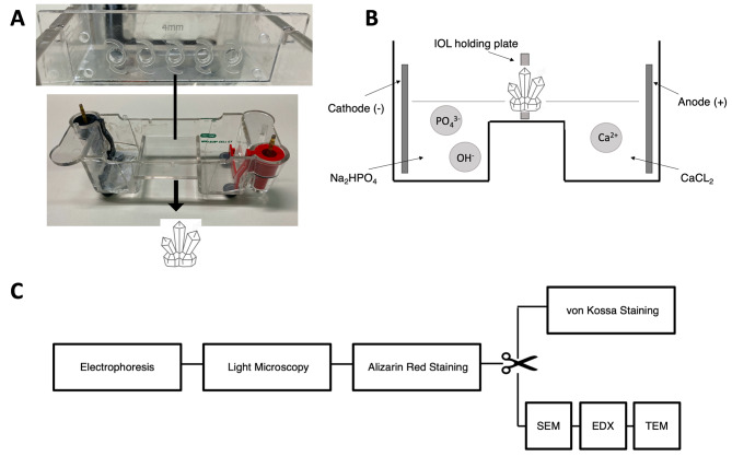
Clinical evidence suggests an association between phosphate concentrations in aqueous humor and the risk of intraocular lens (IOL) calcification. To test this hypothesis the influence of different phosphate concentrations on IOL calcification was evaluated in an in vitro electrophoresis model.

METHODS

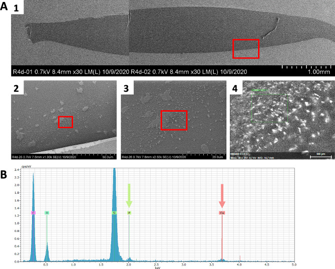
20 IOLs of two hydrophilic IOL models (CT Spheris 204, Zeiss; Lentis L-313, Oculentis) and one hydrophobic control IOL model (Clareon CNA0T0, Alcon) were exposed to physiologic and elevated phosphate concentrations, similar to diabetic aqueous humor. IOL calcification was analyzed by alizarin red staining, von Kossa staining, scanning electron microscopy, energy dispersive x-ray spectroscopy and transmission electron microscopy with electron diffraction.

RESULTS

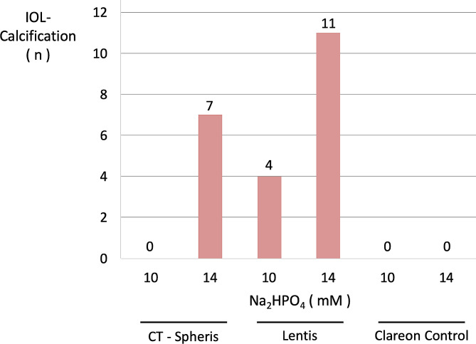
Higher phosphate concentrations were associated with IOL calcification. Analyses of IOL surfaces and cross-sections documented calcification in no CT Spheris and 4 Lentis IOLs following exposure to 10 mM NaHPO, compared with 7 and 11 positive analyses following exposure to 14 mM NaHPO, respectively. Furthermore, a clear association between IOL calcification and the duration of electrophoresis was demonstrated, confirming increased phosphate concentrations and duration of exposure as risk factors of IOL calcification.

CONCLUSIONS

Findings suggest that higher phosphate concentrations in aqueous humor, as seen in diabetic patients, contribute to an increased IOL calcification risk, potentially explaining clinical observations showing an increased risk of IOL calcification in patients with diabetes.

摘要

背景

临床证据表明房水中的磷酸盐浓度与人工晶状体(IOL)钙化的风险之间存在关联。为了验证这一假说,本研究在体外电泳模型中评估了不同磷酸盐浓度对 IOL 钙化的影响。

方法

将两种亲水性 IOL 模型(蔡司 CT Spheris 204;欧科伦特斯 Lentis L-313)的 20 个 IOL 和一个疏水性对照 IOL 模型(爱尔康 Clareon CNA0T0)暴露于生理和升高的磷酸盐浓度下,类似于糖尿病性房水。通过茜素红染色、von Kossa 染色、扫描电子显微镜、能谱分析和电子衍射的透射电子显微镜分析 IOL 钙化。

结果

较高的磷酸盐浓度与 IOL 钙化有关。对 IOL 表面和横截面的分析表明,在暴露于 10 mM NaHPO4 后,4 个 Lentis IOL 和 0 个 CT Spheris IOL 发生钙化,而在暴露于 14 mM NaHPO4 后,分别有 7 个和 11 个分析为阳性。此外,还明确了 IOL 钙化与电泳持续时间之间的关联,证实了较高的磷酸盐浓度和暴露时间是 IOL 钙化的危险因素。

结论

这些发现表明,糖尿病患者房水中较高的磷酸盐浓度可能会增加 IOL 钙化的风险,这可以解释临床上观察到的糖尿病患者 IOL 钙化风险增加的现象。

https://cdn.ncbi.nlm.nih.gov/pmc/blobs/0b52/11342483/edf8b6dbb721/12886_2024_3553_Fig9_HTML.jpg
https://cdn.ncbi.nlm.nih.gov/pmc/blobs/0b52/11342483/7f68a4e36b5e/12886_2024_3553_Fig1_HTML.jpg
https://cdn.ncbi.nlm.nih.gov/pmc/blobs/0b52/11342483/ca9122623b48/12886_2024_3553_Fig2_HTML.jpg
https://cdn.ncbi.nlm.nih.gov/pmc/blobs/0b52/11342483/fb5671e08de1/12886_2024_3553_Fig3_HTML.jpg
https://cdn.ncbi.nlm.nih.gov/pmc/blobs/0b52/11342483/5bd95508716e/12886_2024_3553_Fig4_HTML.jpg
https://cdn.ncbi.nlm.nih.gov/pmc/blobs/0b52/11342483/6bc994edb6ad/12886_2024_3553_Fig5_HTML.jpg
https://cdn.ncbi.nlm.nih.gov/pmc/blobs/0b52/11342483/5cbc0cabc675/12886_2024_3553_Fig6_HTML.jpg
https://cdn.ncbi.nlm.nih.gov/pmc/blobs/0b52/11342483/22882c23e79c/12886_2024_3553_Fig7_HTML.jpg
https://cdn.ncbi.nlm.nih.gov/pmc/blobs/0b52/11342483/248d726ad9cd/12886_2024_3553_Fig8_HTML.jpg
https://cdn.ncbi.nlm.nih.gov/pmc/blobs/0b52/11342483/edf8b6dbb721/12886_2024_3553_Fig9_HTML.jpg

相似文献

1
Higher phosphate concentrations as in aqueous humor of diabetic patients increase intraocular lens calcification.糖尿病患者房水中较高的磷酸盐浓度会增加人工晶状体的钙化。
BMC Ophthalmol. 2024 Aug 23;24(1):363. doi: 10.1186/s12886-024-03553-z.
2
Calcification of Hydroview H60M intraocular lenses: aqueous humor analysis and comparisons with other intraocular lens materials.Hydroview H60M人工晶状体的钙化:房水分析及与其他人工晶状体材料的比较
J Cataract Refract Surg. 2008 Jan;34(1):80-6. doi: 10.1016/j.jcrs.2007.08.020.
3
Development of a standardized in vitro model to reproduce hydrophilic acrylic intraocular lens calcification.开发一种标准化的体外模型以重现亲水性丙烯酸人工晶状体钙化。
Sci Rep. 2022 May 10;12(1):7685. doi: 10.1038/s41598-022-11486-0.
4
Opacification of intraocular lenses implanted during infancy: a clinicopathologic study of 4 explanted intraocular lenses.婴儿期植入的人工晶状体混浊:4 例取出的人工晶状体的临床病理研究。
Ophthalmology. 2011 Nov;118(11):2128-2132.e1. doi: 10.1016/j.ophtha.2011.05.012. Epub 2011 Aug 20.
5
Experimental investigation on mechanism of hydrophilic acrylic intraocular lens calcification.亲水丙烯酸人工晶状体钙化机制的实验研究。
Am J Ophthalmol. 2011 Nov;152(5):824-33.e1. doi: 10.1016/j.ajo.2011.04.009. Epub 2011 Jul 20.
6
[Opacification of a hydrophilic acrylic intraocular lens after DMEK : A material analysis].[DMEK术后亲水性丙烯酸人工晶状体混浊:材料分析]
Ophthalmologe. 2017 Sep;114(9):832-837. doi: 10.1007/s00347-016-0394-6.
7
Hydrophilic acrylic intraocular lens optic opacification in a diabetic patient.糖尿病患者亲水丙烯酸人工晶状体光学混浊。
Jpn J Ophthalmol. 2011 Nov;55(6):595-9. doi: 10.1007/s10384-011-0074-7. Epub 2011 Aug 31.
8
Subsurface calcification of hydrophilic refractive multifocal intraocular lenses with a hydrophobic surface: A case series.具有疏水表面的亲水性折射多焦点人工晶状体的表面下钙化:病例系列
Medicine (Baltimore). 2019 Dec;98(50):e18379. doi: 10.1097/MD.0000000000018379.
9
Calcification of Hydrophilic Acrylic Intraocular Lenses With a Hydrophobic Surface: Laboratory Analysis of 6 Cases.具有疏水表面的亲水性丙烯酸人工晶状体钙化:6例病例的实验室分析
Am J Ophthalmol. 2016 Aug;168:68-77. doi: 10.1016/j.ajo.2016.04.018. Epub 2016 Apr 27.
10
Microscopic Characteristics of Late Intraocular Lens Opacifications.后发性白内障的微观特征。
Arch Pathol Lab Med. 2021 Jun 1;145(6):759-767. doi: 10.5858/arpa.2019-0626-RA.

引用本文的文献

1
How do intraocular lens materials influence the outcome of cataract surgery?人工晶状体材料如何影响白内障手术的结果?
Curr Opin Ophthalmol. 2025 Jan 1;36(1):18-24. doi: 10.1097/ICU.0000000000001095. Epub 2024 Oct 23.

本文引用的文献

1
Development of a standardized in vitro model to reproduce hydrophilic acrylic intraocular lens calcification.开发一种标准化的体外模型以重现亲水性丙烯酸人工晶状体钙化。
Sci Rep. 2022 May 10;12(1):7685. doi: 10.1038/s41598-022-11486-0.
2
[Opacification of intraocular lenses: laboratory and clinical findings].[人工晶状体混浊:实验室及临床研究结果]
Ophthalmologe. 2021 Jul;118(7):633-642. doi: 10.1007/s00347-020-01259-3. Epub 2020 Nov 13.
3
Serial opacification of a hydrophilic-hydrophobic acrylic intraocular lens: analysis of potential risk factors.
亲水性-疏水性丙烯酸眼内晶状体的连续混浊:潜在危险因素分析。
J Cataract Refract Surg. 2020 Dec;46(12):1624-1629. doi: 10.1097/j.jcrs.0000000000000342.
4
Variation in intraocular lens calcification under different environmental conditions in eyes with supplementary sulcus-supported lenses.在植入有辅助性沟支撑人工晶状体的眼中,不同环境条件下人工晶状体钙化的差异。
Am J Ophthalmol Case Rep. 2020 Jun 24;19:100797. doi: 10.1016/j.ajoc.2020.100797. eCollection 2020 Sep.
5
Reasons for explantation, demographics, and material analysis of 200 intraocular lens explants.200 枚人工晶状体取出的原因、人口统计学资料和材料分析。
J Cataract Refract Surg. 2020 Jan;46(1):20-26. doi: 10.1016/j.jcrs.2019.08.045.
6
Impact of Primary Calcification in Segmented Refractive Bifocal Intraocular Lenses on Optical Performance Including Straylight.分段折射双焦点人工晶状体原发性钙化对包括杂散光在内的光学性能的影响。
J Refract Surg. 2020 Jan 1;36(1):20-27. doi: 10.3928/1081597X-20191119-01.
7
Localized calcification of hydrophilic acrylic intraocular lenses after posterior segment procedures.后段手术后继发性亲水丙烯酸人工晶状体局限性钙化。
J Cataract Refract Surg. 2019 Dec;45(12):1801-1807. doi: 10.1016/j.jcrs.2019.07.015.
8
Systematic review of potential causes of intraocular lens opacification.系统评价导致人工晶状体混浊的潜在原因。
Clin Exp Ophthalmol. 2020 Jan;48(1):89-97. doi: 10.1111/ceo.13650. Epub 2019 Oct 22.
9
Cataract in diabetes mellitus.糖尿病性白内障
World J Diabetes. 2019 Mar 15;10(3):140-153. doi: 10.4239/wjd.v10.i3.140.
10
Calcification of hydrophilic acrylic intraocular lenses following secondary surgical procedures in the anterior and posterior segments.眼后段和眼前段二次手术后继发亲水性丙烯酸人工晶状体钙化。
Br J Ophthalmol. 2019 Dec;103(12):1700-1703. doi: 10.1136/bjophthalmol-2018-313385. Epub 2019 Feb 23.