Wu Bing, Zhang Tao, Chen Huabin, Shi Xin, Guan Changbiao, Hu Jianzhong, Lu Hongbin
Department of Sports Medicine, Xiangya Hospital, Central South University, Changsha, 410008, Hunan, China.
Key Laboratory of Organ Injury, Aging and Regenerative Medicine of Hunan Province, Changsha, 410008, Hunan, China.
J Orthop Translat. 2024 Aug 2;48:89-106. doi: 10.1016/j.jot.2024.07.009. eCollection 2024 Sep.
Fibrovascular scar healing of bone-tendon interface (BTI) instead of functional fibrocartilage regeneration is the main concern associated with unsatisfactory prognosis in rotator cuff repair. Mesenchymal stem cells (MSCs) exosomes have been reported to be a new promising cell-free approach for rotator cuff healing. Whereas, controversies abound in whether exosomes of native MSCs alone can effectively induce chondrogenesis.
To explore the effect of exosomes derived from low-intensity pulsed ultrasound stimulation (LIPUS)-preconditioned bone marrow mesenchymal stem cells (LIPUS-BMSC-Exos) or un-preconditioned BMSCs (BMSC-Exos) on rotator cuff healing and the underlying mechanism.
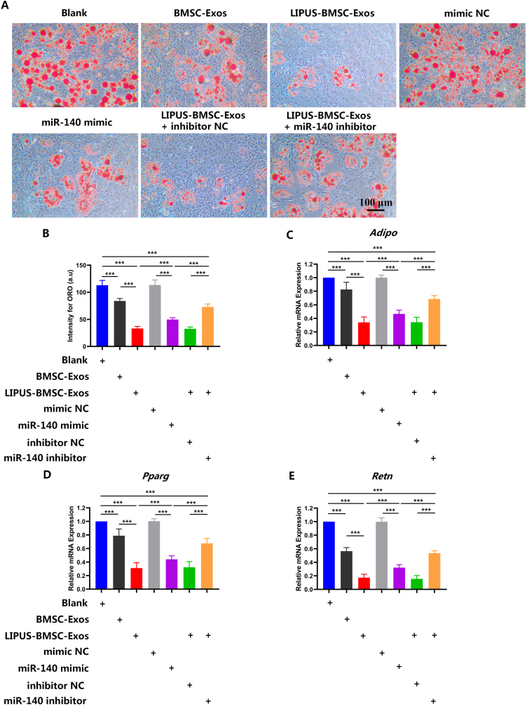
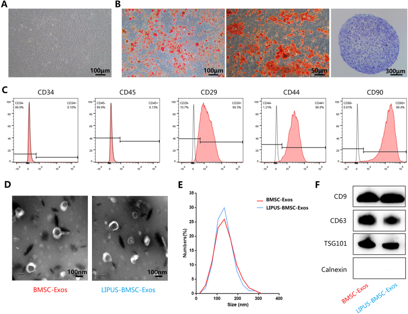
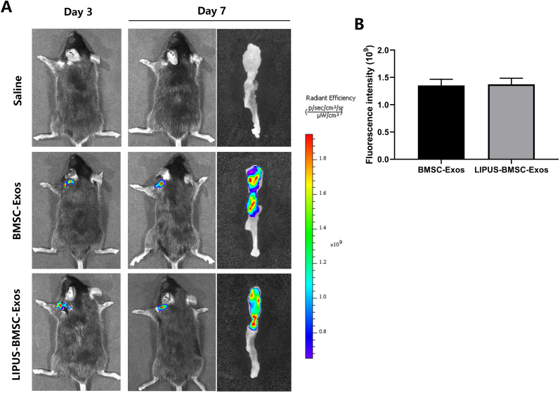
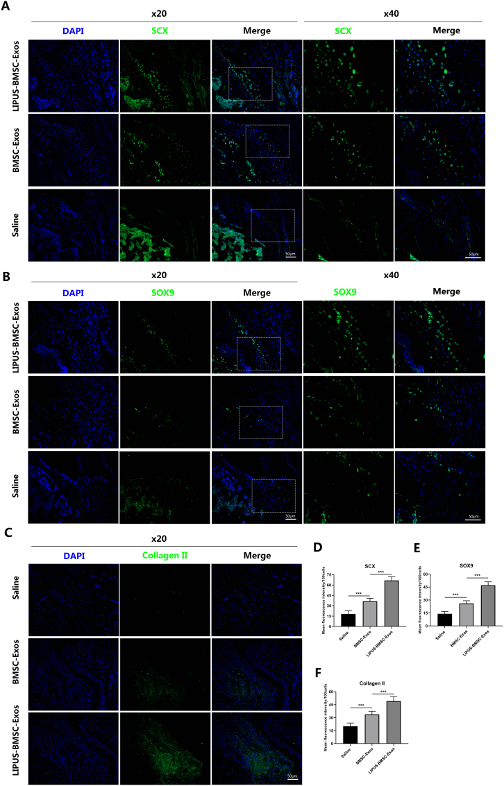
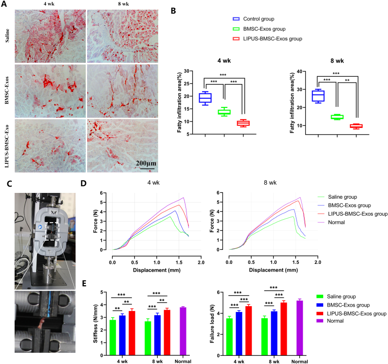
C57BL/6 mice underwent unilateral supraspinatus tendon detachment and repair were randomly assigned to saline, BMSCs-Exos or LIPUS-BMSC-Exos injection therapy. Histological, immunofluorescent and biomechanical tests were detected to investigate the effect of exosomes injection on BTI healing and muscle fatty infiltration of the repaired rotator cuff. , native BMSCs were incubated with BMSC-Exos or LIPUS-BMSC-Exos and then chondrogenic/adipogenic differentiation were observed. Further, quantitative real-time polymerase chain reaction (qRT-PCR) was performed to detect the chondrogenesis/adipogenesis-related miRNA profiles of LIPUS-BMSC-Exos and BMSC-Exos. The chondrogenic/adipogenic potential of the key miRNA was verified through function recover test with its mimic and inhibitor.
The results indicated that the biomechanical properties of the supraspinatus tendon-humeral junction were significantly improved in the LIPUS-BMSC-Exos group than that of the BMSCs-Exos group. The LIPUS-BMSC-Exos group also exhibited a higher histological score and more newly regenerated fibrocartilage at the repair site at postoperative 2 and 4 weeks and less fatty infiltration at 4 weeks than the BMSCs-Exos group. , co-culture of BMSCs with LIPUS-BMSC-Exos could significantly promote BMSCs chondrogenic differentiation and inhibit adipogenic differentiation. Subsequently, qRT-PCR revealed significantly higher enrichment of chondrogenic miRNAs and less enrichment of adipogenic miRNAs in LIPUS-BMSC-Exos compared with BMSC-Exos. Moreover, we demonstrated that this chondrogenesis-inducing potential was primarily attributed to miR-140, one of the most abundant miRNAs in LIPUS-BMSC-Exos.
LIPUS-preconditioned BMSC-Exos can effectively promote BTI fibrocartilage regeneration and ameliorate supraspinatus fatty infiltration by positive regulation of pro-chondrogenesis and anti-adipogenesis, which was primarily through delivering miR-140.
These findings propose an innovative "LIPUS combined Exosomes strategy" for rotator cuff healing which combines both physiotherapeutic and biotherapeutic advantages. This strategy possesses a good translational potential as a local injection of LIPUS preconditioned BMSC-derived Exos during operation can be not only efficient for promoting fibrocartilage regeneration and ameliorating rotator cuff fatty infiltration, but also time-saving, simple and convenient for patients.
肩袖修复预后不理想的主要问题是骨-肌腱界面(BTI)发生纤维血管瘢痕愈合而非功能性纤维软骨再生。间充质干细胞(MSCs)外泌体已被报道是一种用于肩袖愈合的有前景的新型无细胞方法。然而,单纯天然MSCs的外泌体能否有效诱导软骨形成仍存在诸多争议。
探讨低强度脉冲超声刺激预处理的骨髓间充质干细胞(LIPUS-BMSC-Exos)或未预处理的骨髓间充质干细胞(BMSC-Exos)来源的外泌体对肩袖愈合的影响及其潜在机制。
将接受单侧冈上肌腱切断和修复的C57BL/6小鼠随机分为生理盐水组、BMSCs-Exos组或LIPUS-BMSC-Exos注射治疗组。通过组织学、免疫荧光和生物力学测试来研究外泌体注射对BTI愈合及修复后肩袖肌肉脂肪浸润的影响。将天然骨髓间充质干细胞与BMSC-Exos或LIPUS-BMSC-Exos共培养,然后观察软骨形成/脂肪形成分化情况。此外,进行定量实时聚合酶链反应(qRT-PCR)以检测LIPUS-BMSC-Exos和BMSC-Exos中软骨形成/脂肪形成相关的miRNA谱。通过其模拟物和抑制剂的功能恢复试验验证关键miRNA的软骨形成/脂肪形成潜力。
结果表明,LIPUS-BMSC-Exos组冈上肌腱-肱骨交界处的生物力学性能显著优于BMSCs-Exos组。与BMSCs-Exos组相比,LIPUS-BMSC-Exos组在术后2周和4周时修复部位的组织学评分更高,新再生的纤维软骨更多,且在4周时脂肪浸润更少。骨髓间充质干细胞与LIPUS-BMSC-Exos共培养可显著促进骨髓间充质干细胞的软骨形成分化并抑制脂肪形成分化。随后,qRT-PCR显示与BMSC-Exos相比,LIPUS-BMSC-Exos中软骨形成相关miRNA的富集显著更高,脂肪形成相关miRNA的富集更少。此外,我们证明这种软骨形成诱导潜力主要归因于miR-140,它是LIPUS-BMSC-Exos中最丰富的miRNA之一。
LIPUS预处理的BMSC-Exos可通过正向调节促软骨形成和抗脂肪形成,有效促进BTI纤维软骨再生并改善冈上肌脂肪浸润,这主要是通过传递miR-140实现的。
这些发现提出了一种创新的“LIPUS联合外泌体策略”用于肩袖愈合,该策略兼具物理治疗和生物治疗的优势。这种策略具有良好的转化潜力,因为在手术期间局部注射LIPUS预处理的骨髓间充质干细胞来源的外泌体不仅对促进纤维软骨再生和改善肩袖脂肪浸润有效,而且对患者来说省时、简单且方便。