• 文献检索
  • 文档翻译
  • 深度研究
  • 学术资讯
  • Suppr Zotero 插件Zotero 插件
  • 邀请有礼
  • 套餐&价格
  • 历史记录
应用&插件
Suppr Zotero 插件Zotero 插件浏览器插件Mac 客户端Windows 客户端微信小程序
定价
高级版会员购买积分包购买API积分包
服务
文献检索文档翻译深度研究API 文档MCP 服务
关于我们
关于 Suppr公司介绍联系我们用户协议隐私条款
关注我们

Suppr 超能文献

核心技术专利:CN118964589B侵权必究
粤ICP备2023148730 号-1Suppr @ 2026

文献检索

告别复杂PubMed语法,用中文像聊天一样搜索,搜遍4000万医学文献。AI智能推荐,让科研检索更轻松。

立即免费搜索

文件翻译

保留排版,准确专业,支持PDF/Word/PPT等文件格式,支持 12+语言互译。

免费翻译文档

深度研究

AI帮你快速写综述,25分钟生成高质量综述,智能提取关键信息,辅助科研写作。

立即免费体验

多发性硬化症中对比增强病变分割:一种在多中心队列中验证的深度学习方法。

Contrast-Enhancing Lesion Segmentation in Multiple Sclerosis: A Deep Learning Approach Validated in a Multicentric Cohort.

作者信息

Greselin Martina, Lu Po-Jui, Melie-Garcia Lester, Ocampo-Pineda Mario, Galbusera Riccardo, Cagol Alessandro, Weigel Matthias, de Oliveira Siebenborn Nina, Ruberte Esther, Benkert Pascal, Müller Stefanie, Finkener Sebastian, Vehoff Jochen, Disanto Giulio, Findling Oliver, Chan Andrew, Salmen Anke, Pot Caroline, Bridel Claire, Zecca Chiara, Derfuss Tobias, Lieb Johanna M, Diepers Michael, Wagner Franca, Vargas Maria I, Pasquier Renaud Du, Lalive Patrice H, Pravatà Emanuele, Weber Johannes, Gobbi Claudio, Leppert David, Kim Olaf Chan-Hi, Cattin Philippe C, Hoepner Robert, Roth Patrick, Kappos Ludwig, Kuhle Jens, Granziera Cristina

机构信息

Translational Imaging in Neurology (ThINk) Basel, Department of Biomedical Engineering, Faculty of Medicine, University Hospital Basel, University of Basel, 4123 Basel, Switzerland.

Department of Neurology, University Hospital Basel, 4031 Basel, Switzerland.

出版信息

Bioengineering (Basel). 2024 Aug 22;11(8):858. doi: 10.3390/bioengineering11080858.

DOI:10.3390/bioengineering11080858
PMID:39199815
原文链接:https://pmc.ncbi.nlm.nih.gov/articles/PMC11351944/
Abstract

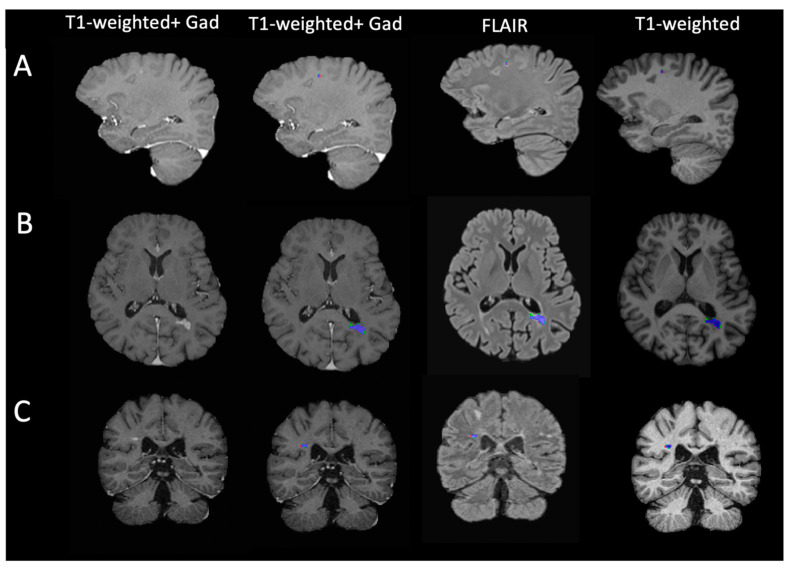
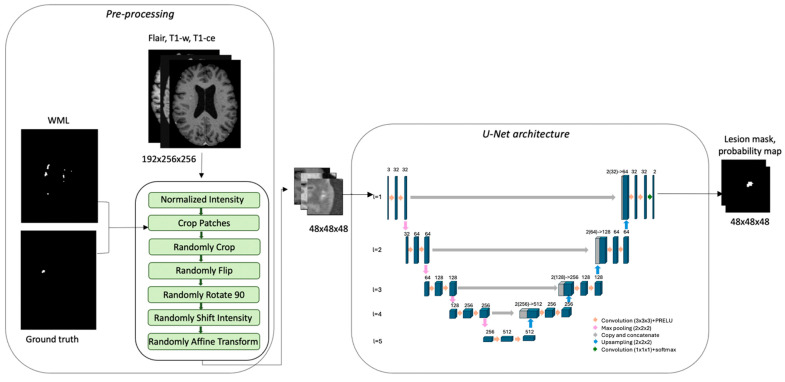
The detection of contrast-enhancing lesions (CELs) is fundamental for the diagnosis and monitoring of patients with multiple sclerosis (MS). This task is time-consuming and suffers from high intra- and inter-rater variability in clinical practice. However, only a few studies proposed automatic approaches for CEL detection. This study aimed to develop a deep learning model that automatically detects and segments CELs in clinical Magnetic Resonance Imaging (MRI) scans. A 3D UNet-based network was trained with clinical MRI from the Swiss Multiple Sclerosis Cohort. The dataset comprised 372 scans from 280 MS patients: 162 showed at least one CEL, while 118 showed no CELs. The input dataset consisted of T1-weighted before and after gadolinium injection, and FLuid Attenuated Inversion Recovery images. The sampling strategy was based on a white matter lesion mask to confirm the existence of real contrast-enhancing lesions. To overcome the dataset imbalance, a weighted loss function was implemented. The Dice Score Coefficient and True Positive and False Positive Rates were 0.76, 0.93, and 0.02, respectively. Based on these results, the model developed in this study might well be considered for clinical decision support.

摘要

检测对比增强病灶(CELs)是多发性硬化症(MS)患者诊断和监测的基础。在临床实践中,这项任务耗时且存在较高的评分者内和评分者间变异性。然而,只有少数研究提出了用于CEL检测的自动方法。本研究旨在开发一种深度学习模型,用于在临床磁共振成像(MRI)扫描中自动检测和分割CELs。基于3D UNet的网络使用来自瑞士多发性硬化症队列的临床MRI进行训练。该数据集包含来自280名MS患者的372次扫描:162次显示至少一个CEL,而118次未显示CEL。输入数据集包括钆注射前后的T1加权图像以及液体衰减反转恢复图像。采样策略基于白质病变掩码以确认真实对比增强病灶的存在。为克服数据集不平衡问题,实施了加权损失函数。Dice评分系数、真阳性率和假阳性率分别为0.76、0.93和0.02。基于这些结果,本研究开发的模型很可能可用于临床决策支持。

https://cdn.ncbi.nlm.nih.gov/pmc/blobs/aec2/11351944/22b158a1bcde/bioengineering-11-00858-g003.jpg
https://cdn.ncbi.nlm.nih.gov/pmc/blobs/aec2/11351944/5c20672f3ac9/bioengineering-11-00858-g001.jpg
https://cdn.ncbi.nlm.nih.gov/pmc/blobs/aec2/11351944/db61aaeb624b/bioengineering-11-00858-g002.jpg
https://cdn.ncbi.nlm.nih.gov/pmc/blobs/aec2/11351944/22b158a1bcde/bioengineering-11-00858-g003.jpg
https://cdn.ncbi.nlm.nih.gov/pmc/blobs/aec2/11351944/5c20672f3ac9/bioengineering-11-00858-g001.jpg
https://cdn.ncbi.nlm.nih.gov/pmc/blobs/aec2/11351944/db61aaeb624b/bioengineering-11-00858-g002.jpg
https://cdn.ncbi.nlm.nih.gov/pmc/blobs/aec2/11351944/22b158a1bcde/bioengineering-11-00858-g003.jpg

相似文献

1
Contrast-Enhancing Lesion Segmentation in Multiple Sclerosis: A Deep Learning Approach Validated in a Multicentric Cohort.多发性硬化症中对比增强病变分割:一种在多中心队列中验证的深度学习方法。
Bioengineering (Basel). 2024 Aug 22;11(8):858. doi: 10.3390/bioengineering11080858.
2
2.5D transfer deep learning model for segmentation of contrast-enhancing lesions on brain magnetic resonance imaging of multiple sclerosis and neuromyelitis optica spectrum disorder.用于多发性硬化症和视神经脊髓炎谱系障碍脑磁共振成像中强化病变分割的2.5D转移深度学习模型
Quant Imaging Med Surg. 2024 Jan 3;14(1):273-290. doi: 10.21037/qims-23-846. Epub 2023 Nov 15.
3
Deep learning segmentation of gadolinium-enhancing lesions in multiple sclerosis.深度学习对多发性硬化症钆增强病变的分割。
Mult Scler. 2021 Apr;27(4):519-527. doi: 10.1177/1352458520921364. Epub 2020 May 22.
4
Joint MRI T1 Unenhancing and Contrast-enhancing Multiple Sclerosis Lesion Segmentation with Deep Learning in OPERA Trials.基于 OPERA 试验的深度学习方法对 MRI T1 未增强相与增强相多发性硬化病变的分割。
Radiology. 2022 Mar;302(3):662-673. doi: 10.1148/radiol.211528. Epub 2021 Dec 14.
5
Automatic segmentation of gadolinium-enhancing lesions in multiple sclerosis using deep learning from clinical MRI.基于临床 MRI 的深度学习对多发性硬化钆增强病灶的自动分割。
PLoS One. 2021 Sep 1;16(9):e0255939. doi: 10.1371/journal.pone.0255939. eCollection 2021.
6
Automatic segmentation of white matter hyperintensities: validation and comparison with state-of-the-art methods on both Multiple Sclerosis and elderly subjects.自动分割脑白质高信号:在多发性硬化症和老年患者中验证和比较最先进的方法。
Neuroimage Clin. 2022;33:102940. doi: 10.1016/j.nicl.2022.102940. Epub 2022 Jan 10.
7
Are multi-contrast magnetic resonance images necessary for segmenting multiple sclerosis brains? A large cohort study based on deep learning.多对比度磁共振成像对于多发性硬化症大脑的分割是否必要?一项基于深度学习的大型队列研究。
Magn Reson Imaging. 2020 Jan;65:8-14. doi: 10.1016/j.mri.2019.10.003. Epub 2019 Oct 25.
8
Improving the detection of new lesions in multiple sclerosis with a cascaded 3D fully convolutional neural network approach.采用级联3D全卷积神经网络方法提高多发性硬化症新病灶的检测能力。
Front Neurosci. 2022 Nov 24;16:1007619. doi: 10.3389/fnins.2022.1007619. eCollection 2022.
9
Automatic intraprostatic lesion segmentation in multiparametric magnetic resonance images with proposed multiple branch UNet.利用提出的多分支U-Net在多参数磁共振图像中实现前列腺内病变的自动分割。
Med Phys. 2020 Dec;47(12):6421-6429. doi: 10.1002/mp.14517. Epub 2020 Oct 24.
10
Fully automated longitudinal segmentation of new or enlarged multiple sclerosis lesions using 3D convolutional neural networks.使用 3D 卷积神经网络全自动纵向分割新的或扩大的多发性硬化病变。
Neuroimage Clin. 2020;28:102445. doi: 10.1016/j.nicl.2020.102445. Epub 2020 Sep 24.

引用本文的文献

1
Mitigating catastrophic forgetting in Multiple sclerosis lesion segmentation using elastic weight consolidation.使用弹性权重巩固减轻多发性硬化症病变分割中的灾难性遗忘
Neuroimage Clin. 2025;46:103795. doi: 10.1016/j.nicl.2025.103795. Epub 2025 May 20.

本文引用的文献

1
2.5D transfer deep learning model for segmentation of contrast-enhancing lesions on brain magnetic resonance imaging of multiple sclerosis and neuromyelitis optica spectrum disorder.用于多发性硬化症和视神经脊髓炎谱系障碍脑磁共振成像中强化病变分割的2.5D转移深度学习模型
Quant Imaging Med Surg. 2024 Jan 3;14(1):273-290. doi: 10.21037/qims-23-846. Epub 2023 Nov 15.
2
3D U-Net for automated detection of multiple sclerosis lesions: utility of transfer learning from other pathologies.用于自动检测多发性硬化症病变的3D U-Net:从其他病理学进行迁移学习的效用。
Front Neurosci. 2023 Oct 27;17:1188336. doi: 10.3389/fnins.2023.1188336. eCollection 2023.
3
AI-based detection of contrast-enhancing MRI lesions in patients with multiple sclerosis.
基于人工智能检测多发性硬化症患者的磁共振成像对比增强病变。
Insights Imaging. 2023 Jul 16;14(1):123. doi: 10.1186/s13244-023-01460-3.
4
Feature preserving mesh network for semantic segmentation of retinal vasculature to support ophthalmic disease analysis.用于视网膜血管语义分割以支持眼科疾病分析的特征保留网格网络。
Front Med (Lausanne). 2023 Jan 13;9:1040562. doi: 10.3389/fmed.2022.1040562. eCollection 2022.
5
Self-supervised learning in medicine and healthcare.医学和医疗保健中的自我监督学习。
Nat Biomed Eng. 2022 Dec;6(12):1346-1352. doi: 10.1038/s41551-022-00914-1. Epub 2022 Aug 11.
6
Nonlesional Sources of Contrast Enhancement on Postgadolinium "Black-Blood" 3D T1-SPACE Images in Patients with Multiple Sclerosis.多发性硬化症患者钆后“黑血”3D T1-SPACE 图像上非病灶性对比增强源。
AJNR Am J Neuroradiol. 2022 Jun;43(6):872-880. doi: 10.3174/ajnr.A7529. Epub 2022 May 26.
7
Multiple sclerosis cortical lesion detection with deep learning at ultra-high-field MRI.深度学习在超高场 MRI 下对多发性硬化皮质病变的检测。
NMR Biomed. 2022 Aug;35(8):e4730. doi: 10.1002/nbm.4730. Epub 2022 Mar 31.
8
Joint MRI T1 Unenhancing and Contrast-enhancing Multiple Sclerosis Lesion Segmentation with Deep Learning in OPERA Trials.基于 OPERA 试验的深度学习方法对 MRI T1 未增强相与增强相多发性硬化病变的分割。
Radiology. 2022 Mar;302(3):662-673. doi: 10.1148/radiol.211528. Epub 2021 Dec 14.
9
Automatic segmentation of gadolinium-enhancing lesions in multiple sclerosis using deep learning from clinical MRI.基于临床 MRI 的深度学习对多发性硬化钆增强病灶的自动分割。
PLoS One. 2021 Sep 1;16(9):e0255939. doi: 10.1371/journal.pone.0255939. eCollection 2021.
10
Redefining use of MRI for patients with multiple sclerosis.重新定义磁共振成像在多发性硬化症患者中的应用。
Lancet Neurol. 2021 Aug;20(8):591-592. doi: 10.1016/S1474-4422(21)00203-9.