Łobacz Michał, Wieczorek Katarzyna, Mertowska Paulina, Mertowski Sebastian, Kos Marek, Grywalska Ewelina, Hajduk Grzegorz, Rahnama-Hezavah Mansur
Chair and Department of Oral Surgery, Medical University of Lublin, 20-093 Lublin, Poland.
Department of Experimental Immunology, Medical University of Lublin, 20-093 Lublin, Poland.
J Clin Med. 2024 Aug 8;13(16):4638. doi: 10.3390/jcm13164638.
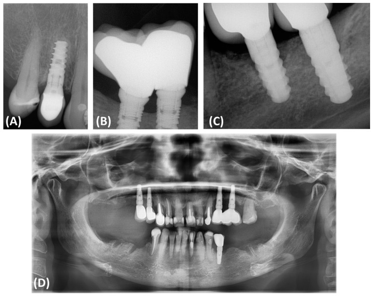
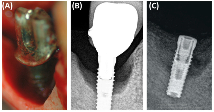
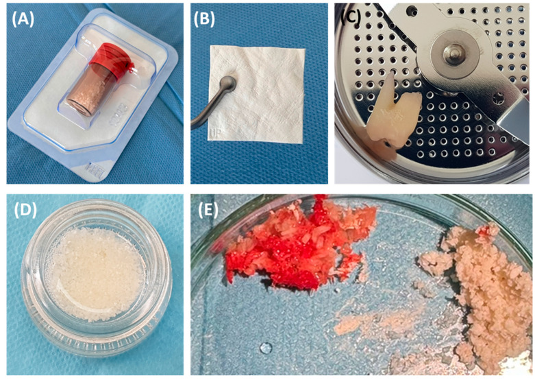
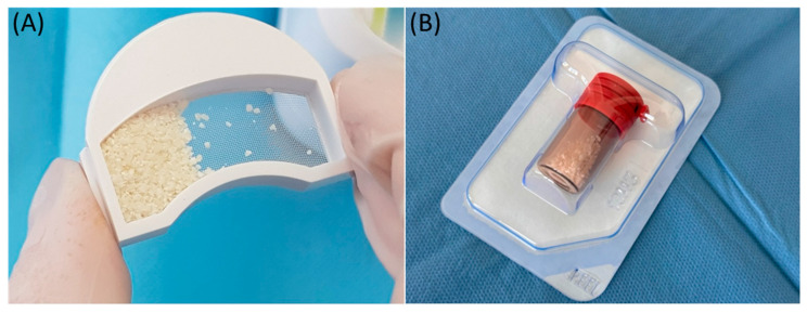
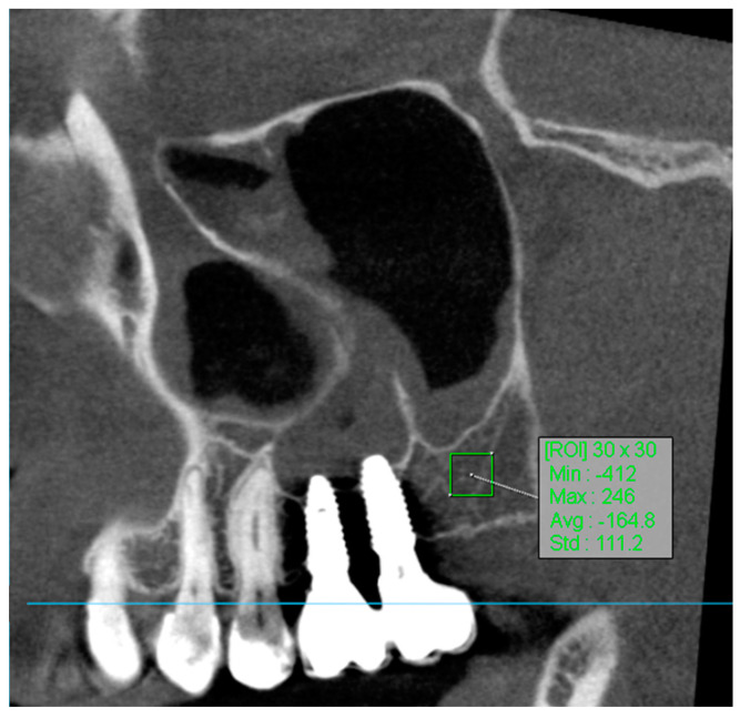
Peri-implantitis is a serious complication in dental implantology that, if left untreated, may lead to implant loss and systemic diseases. Effective regeneration of bone defects resulting from peri-implantitis is crucial to maintaining the functionality of dental implants. The study aimed to compare the effectiveness of fine-particle dentin and Bio-Oss in the reconstruction of bone defects caused by peri-implantitis. The study included a comprehensive radiological assessment of changes in bone density over time. Bone density was assessed using Hounsfield Units (HUs) as a measure of bone attenuation, with radiological assessments performed at 8- and 12-week intervals during the healing process. The study included participants ranging in age from 30 to 65 years. Fifty-seven patients were divided into three groups: 22 patients received small-particle dentin, 15 received Bio-Oss, and 20 controls without bone substitute material. The fine-dentin group showed a 20% increase in bone density after 8 weeks ( < 0.05), while the Bio-Oss group showed a 15% increase after 12 weeks ( < 0.05). The control group showed minimal changes in bone density (5% after 12 weeks), which was not statistically significant. Clinical evaluations showed 95% successful integration in the fine dentin group, 85% in the Bio-Oss group, and 70% in the control group. The fine-dentin group showed a 20% increase in bone density after 8 weeks ( < 0.05), while the Bio-Oss group showed a 15% increase after 12 weeks ( < 0.05). The control group showed minimal changes in bone density (5% after 12 weeks), which was not statistically significant. Clinical evaluations showed 95% successful integration in the fine-dentin group, 85% in the Bio-Oss group, and 70% in the control group. : Both fine-particle dentin and Bio-Oss significantly improved bone density compared to the control group. Fine-particle dentin is suitable for immediate bone regeneration due to its rapid initial regeneration, while Bio-Oss provides long-term support, ideal for maintaining implant stability over a longer period of time. The results highlight the importance of selecting appropriate bone replacement materials depending on the clinical scenario to improve patient outcomes after dental implant placement.
种植体周围炎是牙种植学中的一种严重并发症,若不治疗,可能导致种植体脱落和全身性疾病。有效修复种植体周围炎导致的骨缺损对于维持牙种植体的功能至关重要。本研究旨在比较细颗粒牙本质和Bio-Oss在修复种植体周围炎所致骨缺损方面的有效性。该研究包括对骨密度随时间变化的全面影像学评估。使用亨氏单位(HUs)评估骨密度,作为骨衰减的指标,在愈合过程中每隔8周和12周进行影像学评估。该研究纳入了年龄在30至65岁之间的参与者。57名患者被分为三组:22名患者接受小颗粒牙本质,15名接受Bio-Oss,20名作为未使用骨替代材料的对照组。细颗粒牙本质组在8周后骨密度增加了20%(P<0.05),而Bio-Oss组在12周后增加了15%(P<0.05)。对照组骨密度变化极小(12周后为5%),无统计学意义。临床评估显示,细颗粒牙本质组的成功整合率为95%,Bio-Oss组为85%,对照组为70%。细颗粒牙本质组在8周后骨密度增加了20%(P<0.05),而Bio-Oss组在12周后增加了15%(P<0.05)。对照组骨密度变化极小(12周后为5%),无统计学意义。临床评估显示,细颗粒牙本质组的成功整合率为95%,Bio-Oss组为85%,对照组为70%。与对照组相比,细颗粒牙本质和Bio-Oss均显著提高了骨密度。细颗粒牙本质因其快速的初始再生适合即刻骨再生,而Bio-Oss提供长期支持,非常适合在较长时间内维持种植体稳定性。结果突出了根据临床情况选择合适的骨替代材料以改善牙种植术后患者预后的重要性。