Department of Ophthalmology, Hallym University Sacred Heart Hospital, Anyang, Republic of Korea.
Casey Eye Institute, Oculofacial Plastic and Reconstructive Surgery, Casey Aesthetic Facial Surgery Center, Oregon Health & Science University, Portland, OR, United States of America.
PLoS One. 2024 Aug 30;19(8):e0308528. doi: 10.1371/journal.pone.0308528. eCollection 2024.
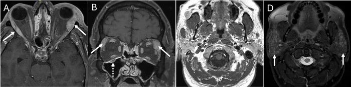
This study investigates the accuracy of either computerized tomography (CT) or magnetic resonance imaging (MRI) for the evaluation of various orbital diseases.
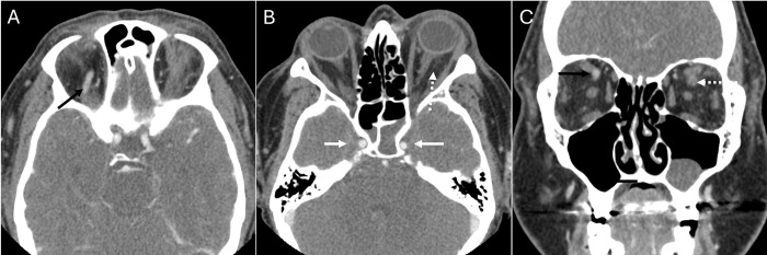
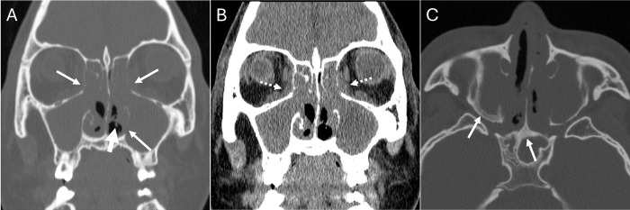
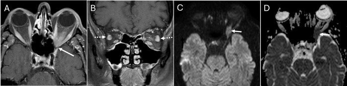
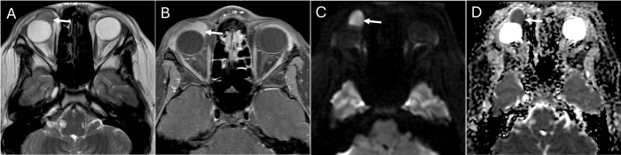
We collected 126 CT scans and 65 MRI scans from 144 subjects and asked two radiologists to interpret the images without clinical information. Images included 14 with a clinical diagnosis of orbital infection, 144 with orbital inflammation, and 33 with orbital neoplasm. The inflammatory diseases included thyroid eye disease (TED, n = 69), non-specific orbital inflammation (NSOI, n = 44), IgG4-related disease (IgG4-RD, n = 15), sarcoidosis (Sarcoid, n = 9), granulomatosis with polyangiitis (GPA, n = 5), and Erdheim-Chester disease (ECD, n = 2).
The balanced accuracy (BA) for the two radiologists ranged from 0.87 to 0.90 for cellulitis, 0.81 to 0.86 for inflammation, and 0.82 to 0.85 for neoplasm. Radiologists were excellent at recognizing GPA (BA = 0.98 to 0.99) and very good for TED (BA = 0.80 to 0.86). They also did well identifying IgG4-RD (BA = 0.75 to 0.77), but slightly less well for NSOI (BA = 0.69 to 0.75) and poorly for Sarcoid (BA = 0.48 to 0.50).
CT or MRI scanning contributes to the evaluation of patients with orbital disease, but accuracy does varies based depending on the diagnosis. We could not evaluate issues such as determination of disease activity, variability based on the unit used for imaging or the skills beyond those of our two specialized neuroradiologists. Future studies should directly compare the two imaging modalities and assess the utility of imaging to determine disease activity.
本研究旨在评估计算机断层扫描(CT)或磁共振成像(MRI)在各种眼眶疾病评估中的准确性。
我们收集了 144 名患者的 126 次 CT 扫描和 65 次 MRI 扫描,并要求两名放射科医生在不了解临床信息的情况下对图像进行解读。这些图像包括 14 例临床诊断为眼眶感染,144 例眼眶炎症和 33 例眼眶肿瘤。炎症性疾病包括甲状腺眼病(TED,n=69)、非特异性眼眶炎症(NSOI,n=44)、IgG4 相关疾病(IgG4-RD,n=15)、结节病(Sarcoid,n=9)、肉芽肿性多血管炎(GPA,n=5)和 Erdheim-Chester 病(ECD,n=2)。
两名放射科医生的平衡准确率(BA)在蜂窝织炎为 0.87 至 0.90,炎症为 0.81 至 0.86,肿瘤为 0.82 至 0.85。放射科医生非常擅长识别 GPA(BA=0.98 至 0.99)和 TED(BA=0.80 至 0.86)。他们还能很好地识别 IgG4-RD(BA=0.75 至 0.77),但对 NSOI(BA=0.69 至 0.75)的识别能力稍差,对 Sarcoid(BA=0.48 至 0.50)的识别能力较差。
CT 或 MRI 扫描有助于评估眼眶疾病患者,但准确性因诊断而异。我们无法评估疾病活动的确定、成像使用的单位的变化或超出我们两名神经放射科医生技能的问题。未来的研究应直接比较两种成像方式,并评估成像在确定疾病活动方面的效用。