, Florence, Italy.
Department of Experimental and Clinical Medicine, University of Florence, 50134, Florence, Italy.
Radiol Med. 2024 Nov;129(11):1682-1695. doi: 10.1007/s11547-024-01882-z. Epub 2024 Sep 3.
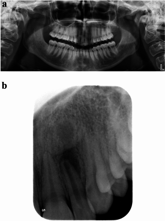
Apical periodontitis (AP) is one of the most common pathologies of the oral cavity. An early and accurate diagnosis of AP lesions is crucial for proper management and planning of endodontic treatments. This study investigated the diagnostic accuracy of periapical radiography (PR) and panoramic radiography (PAN) in the detection of clinically/surgically/histopathologically confirmed AP lesions.
A systematic literature review was conducted in accordance with the PRISMA guidelines. The search strategy was limited to English language articles via PubMed, Embase and Web of Science databases up to June 30, 2023. Such articles provided diagnostic accuracy values of PR and/or PAN in the detection of AP lesions or alternatively data needed to calculate them.
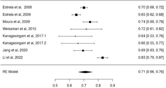
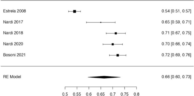
Twelve studies met inclusion criteria and were considered for the analysis. The average value of diagnostic accuracy in assessing AP lesions was 71% for PR and 66% for PAN. According to different accuracy for specific anatomical areas, it is recommended to use PR in the analysis of AP lesions located in the upper arch and lower incisor area, whereas lower premolar and molar areas may be investigated with the same accuracy with PR or PAN.
Two-dimensional imaging must be considered the first-level examination for the diagnosis of AP lesions. PR had an overall slightly higher diagnostic accuracy than PAN. Evidence from this review provided a useful tool to support radiologists and dentists in their decision-making when inflammatory periapical bone lesions are suspected to achieve the best clinical outcome for patients, improving the quality of clinical practice.
根尖周炎(AP)是口腔最常见的疾病之一。早期准确诊断 AP 病变对于根管治疗的适当管理和计划至关重要。本研究旨在探讨根尖片(PR)和全景片(PAN)在检测临床/手术/组织病理学证实的 AP 病变中的诊断准确性。
根据 PRISMA 指南进行系统文献回顾。检索策略仅限于英语文章,通过 PubMed、Embase 和 Web of Science 数据库检索至 2023 年 6 月 30 日。这些文章提供了 PR 和/或 PAN 检测 AP 病变的诊断准确性值,或者提供了计算这些值所需的数据。
12 项研究符合纳入标准并进行了分析。PR 评估 AP 病变的平均诊断准确性值为 71%,PAN 为 66%。根据特定解剖区域的不同准确性,建议在上颌弓和下切牙区域的 AP 病变分析中使用 PR,而下前磨牙和磨牙区域可以使用 PR 或 PAN 以相同的准确性进行研究。
二维成像必须被视为诊断 AP 病变的首选检查方法。PR 的总体诊断准确性略高于 PAN。本综述提供的证据为放射科医生和牙医在怀疑存在炎症性根尖骨病变时做出决策提供了有用的工具,以实现患者的最佳临床结果,提高临床实践质量。