• 文献检索
  • 文档翻译
  • 深度研究
  • 学术资讯
  • Suppr Zotero 插件Zotero 插件
  • 邀请有礼
  • 套餐&价格
  • 历史记录
应用&插件
Suppr Zotero 插件Zotero 插件浏览器插件Mac 客户端Windows 客户端微信小程序
定价
高级版会员购买积分包购买API积分包
服务
文献检索文档翻译深度研究API 文档MCP 服务
关于我们
关于 Suppr公司介绍联系我们用户协议隐私条款
关注我们

Suppr 超能文献

核心技术专利:CN118964589B侵权必究
粤ICP备2023148730 号-1Suppr @ 2026

文献检索

告别复杂PubMed语法,用中文像聊天一样搜索,搜遍4000万医学文献。AI智能推荐,让科研检索更轻松。

立即免费搜索

文件翻译

保留排版,准确专业,支持PDF/Word/PPT等文件格式,支持 12+语言互译。

免费翻译文档

深度研究

AI帮你快速写综述,25分钟生成高质量综述,智能提取关键信息,辅助科研写作。

立即免费体验

深度学习驱动的无标记活细胞染色质敏感部分波谱显微镜成像的自动细胞核分割

Deep Learning-driven Automatic Nuclei Segmentation of Label-free Live Cell Chromatin-sensitive Partial Wave Spectroscopic Microscopy Imaging.

作者信息

Alom Shahin, Daneshkhah Ali, Acosta Nicolas, Anthony Nick, Liwag Emily Pujadas, Backman Vadim, Gaire Sunil Kumar

机构信息

Department of Electrical and Computer Engineering, North Carolina Agricultural and Technical State University, Greensboro, NC 27411, USA.

Department of Biomedical Engineering, Northwestern University, Evanston, IL 60208, USA.

出版信息

bioRxiv. 2024 Aug 21:2024.08.20.608885. doi: 10.1101/2024.08.20.608885.

DOI:10.1101/2024.08.20.608885
PMID:39229026
原文链接:https://pmc.ncbi.nlm.nih.gov/articles/PMC11370422/
Abstract

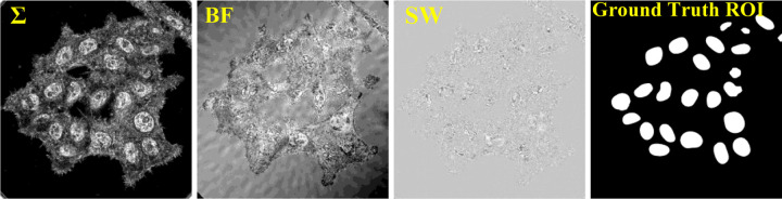
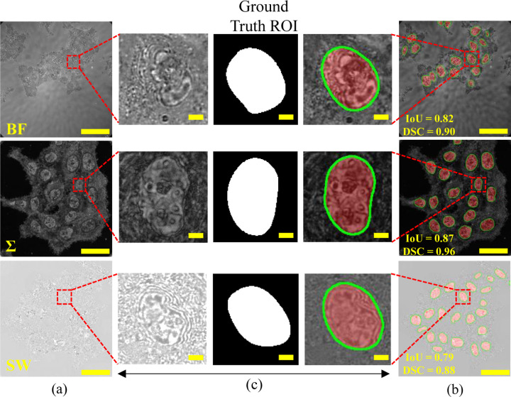
Chromatin-sensitive Partial Wave Spectroscopic (csPWS) microscopy offers a non-invasive glimpse into the mass density distribution of cellular structures at the nanoscale, leveraging the spectroscopic information. Such capability allows us to analyze the chromatin structure and organization and the global transcriptional state of the cell nuclei for the study of its role in carcinogenesis. Accurate segmentation of the nuclei in csPWS microscopy images is an essential step in isolating them for further analysis. However, manual segmentation is error-prone, biased, time-consuming, and laborious, resulting in disrupted nuclear boundaries with partial or over-segmentation. Here, we present an innovative deep-learning-driven approach to automate the accurate nuclei segmentation of label-free live cell csPWS microscopy imaging data. Our approach, csPWS-seg, harnesses the Convolutional Neural Networks-based U-Net model with an attention mechanism to automate the accurate cell nuclei segmentation of csPWS microscopy images. We leveraged the structural, physical, and biological differences between the cytoplasm, nucleus, and nuclear periphery to construct three distinct csPWS feature images for nucleus segmentation. Using these images of HCT116 cells, csPWS-seg achieved superior performance with a median Intersection over Union (IoU) of 0.80 and a Dice Similarity Coefficient (DSC) score of 0.88. The csPWS-seg overcame the segmentation performance over the baseline U-Net model and another attention-based model, SE-U-Net, marking a significant improvement in segmentation accuracy. Further, we analyzed the performance of our proposed model with four loss functions: binary cross-entropy loss, focal loss, dice loss, and Jaccard loss. The csPWS-seg with focal loss provided the best results compared to other loss functions. The automatic and accurate nuclei segmentation offered by the csPWS-seg not only automates, accelerates, and streamlines csPWS data analysis but also enhances the reliability of subsequent chromatin analysis research, paving the way for more accurate diagnostics, treatment, and understanding of cellular mechanisms for carcinogenesis.

摘要

染色质敏感的部分波谱显微镜(csPWS)利用光谱信息,能够对纳米尺度下细胞结构的质量密度分布进行非侵入性观察。这种能力使我们能够分析染色质的结构和组织以及细胞核的整体转录状态,以研究其在致癌过程中的作用。在csPWS显微镜图像中准确分割细胞核是将其分离出来进行进一步分析的关键步骤。然而,手动分割容易出错、存在偏差、耗时且费力,会导致核边界中断,出现部分分割或过度分割的情况。在此,我们提出一种创新的深度学习驱动方法,用于自动准确分割无标记活细胞csPWS显微镜成像数据中的细胞核。我们的方法csPWS-seg利用基于卷积神经网络的U-Net模型并结合注意力机制,自动准确分割csPWS显微镜图像中的细胞核。我们利用细胞质、细胞核和核周之间的结构、物理和生物学差异,构建了三张不同的用于细胞核分割的csPWS特征图像。使用这些HCT116细胞的图像,csPWS-seg表现出色,平均交并比(IoU)为0.80,骰子相似系数(DSC)得分为0.88。csPWS-seg在分割性能上超过了基线U-Net模型和另一个基于注意力的模型SE-U-Net,在分割精度上有显著提高。此外,我们用四种损失函数分析了我们提出的模型的性能:二元交叉熵损失、焦点损失、骰子损失和杰卡德损失。与其他损失函数相比,采用焦点损失的csPWS-seg提供了最佳结果。csPWS-seg提供的自动且准确的细胞核分割不仅使csPWS数据分析自动化、加速并简化,还提高了后续染色质分析研究的可靠性,为更准确的诊断、治疗以及理解致癌的细胞机制铺平了道路。

https://cdn.ncbi.nlm.nih.gov/pmc/blobs/e747/11370422/75ea37908c1f/nihpp-2024.08.20.608885v1-f0010.jpg
https://cdn.ncbi.nlm.nih.gov/pmc/blobs/e747/11370422/5eccc912e547/nihpp-2024.08.20.608885v1-f0001.jpg
https://cdn.ncbi.nlm.nih.gov/pmc/blobs/e747/11370422/ec9bbe41dac9/nihpp-2024.08.20.608885v1-f0002.jpg
https://cdn.ncbi.nlm.nih.gov/pmc/blobs/e747/11370422/7ff48839a823/nihpp-2024.08.20.608885v1-f0003.jpg
https://cdn.ncbi.nlm.nih.gov/pmc/blobs/e747/11370422/dd1666fd126e/nihpp-2024.08.20.608885v1-f0004.jpg
https://cdn.ncbi.nlm.nih.gov/pmc/blobs/e747/11370422/001bc8e3b39c/nihpp-2024.08.20.608885v1-f0005.jpg
https://cdn.ncbi.nlm.nih.gov/pmc/blobs/e747/11370422/8041dbc7ca46/nihpp-2024.08.20.608885v1-f0006.jpg
https://cdn.ncbi.nlm.nih.gov/pmc/blobs/e747/11370422/150d0850df4b/nihpp-2024.08.20.608885v1-f0007.jpg
https://cdn.ncbi.nlm.nih.gov/pmc/blobs/e747/11370422/3b129c1181ca/nihpp-2024.08.20.608885v1-f0008.jpg
https://cdn.ncbi.nlm.nih.gov/pmc/blobs/e747/11370422/35fb1a914e4f/nihpp-2024.08.20.608885v1-f0009.jpg
https://cdn.ncbi.nlm.nih.gov/pmc/blobs/e747/11370422/75ea37908c1f/nihpp-2024.08.20.608885v1-f0010.jpg

相似文献

1
Deep Learning-driven Automatic Nuclei Segmentation of Label-free Live Cell Chromatin-sensitive Partial Wave Spectroscopic Microscopy Imaging.深度学习驱动的无标记活细胞染色质敏感部分波谱显微镜成像的自动细胞核分割
bioRxiv. 2024 Aug 21:2024.08.20.608885. doi: 10.1101/2024.08.20.608885.
2
Focus U-Net: A novel dual attention-gated CNN for polyp segmentation during colonoscopy.聚焦 U-Net:一种新颖的双注意力门控 CNN,用于结肠镜检查中的息肉分割。
Comput Biol Med. 2021 Oct;137:104815. doi: 10.1016/j.compbiomed.2021.104815. Epub 2021 Sep 2.
3
Automatic prostate segmentation using deep learning on clinically diverse 3D transrectal ultrasound images.基于临床多样的三维经直肠超声图像,利用深度学习进行前列腺自动分割。
Med Phys. 2020 Jun;47(6):2413-2426. doi: 10.1002/mp.14134. Epub 2020 Apr 8.
4
3D Attention M-net for Short-axis Left Ventricular Myocardium Segmentation in Mice MR cardiac Images.3D Attention M-net 用于小鼠 MR 心脏图像的短轴左心室心肌分割。
Annu Int Conf IEEE Eng Med Biol Soc. 2021 Nov;2021:3353-3357. doi: 10.1109/EMBC46164.2021.9630335.
5
Dense-UNet: a novel multiphoton cellular image segmentation model based on a convolutional neural network.密集型U-Net:一种基于卷积神经网络的新型多光子细胞图像分割模型。
Quant Imaging Med Surg. 2020 Jun;10(6):1275-1285. doi: 10.21037/qims-19-1090.
6
Automatic Pancreatic Cyst Lesion Segmentation on EUS Images Using a Deep-Learning Approach.基于深度学习的超声内镜图像胰腺囊性病变自动分割。
Sensors (Basel). 2021 Dec 30;22(1):245. doi: 10.3390/s22010245.
7
Improved automatic segmentation of brain metastasis gross tumor volume in computed tomography images for radiotherapy: a position attention module for U-Net architecture.用于放疗的计算机断层扫描图像中脑转移瘤大体肿瘤体积的改进自动分割:一种用于U-Net架构的位置注意力模块
Quant Imaging Med Surg. 2024 Jul 1;14(7):4475-4489. doi: 10.21037/qims-23-1627. Epub 2024 May 24.
8
Layered deep learning for automatic mandibular segmentation in cone-beam computed tomography.基于分层深度学习的锥形束计算机断层扫描下颌骨自动分割。
J Dent. 2021 Nov;114:103786. doi: 10.1016/j.jdent.2021.103786. Epub 2021 Aug 20.
9
Densely Convolutional Spatial Attention Network for nuclei segmentation of histological images for computational pathology.用于计算病理学组织学图像细胞核分割的密集卷积空间注意力网络。
Front Oncol. 2023 May 25;13:1009681. doi: 10.3389/fonc.2023.1009681. eCollection 2023.
10
Effect of learning parameters on the performance of the U-Net architecture for cell nuclei segmentation from microscopic cell images.学习参数对基于 U-Net 架构的微观细胞图像中细胞核分割性能的影响。
Microscopy (Oxf). 2023 Jun 8;72(3):249-264. doi: 10.1093/jmicro/dfac063.

本文引用的文献

1
Machine vision-based autonomous road hazard avoidance system for self-driving vehicles.用于自动驾驶车辆的基于机器视觉的自主道路危险规避系统。
Sci Rep. 2024 May 28;14(1):12178. doi: 10.1038/s41598-024-62629-4.
2
Deep learning-based spectroscopic single-molecule localization microscopy.基于深度学习的光谱单分子定位显微镜。
J Biomed Opt. 2024 Jun;29(6):066501. doi: 10.1117/1.JBO.29.6.066501. Epub 2024 May 24.
3
Early screening of colorectal cancer using feature engineering with artificial intelligence-enhanced analysis of nanoscale chromatin modifications.
利用人工智能增强的纳米级染色质修饰特征工程进行结直肠癌早期筛查。
Sci Rep. 2024 Apr 2;14(1):7808. doi: 10.1038/s41598-024-58016-8.
4
A cognitive deep learning approach for medical image processing.一种用于医学图像处理的认知深度学习方法。
Sci Rep. 2024 Feb 24;14(1):4539. doi: 10.1038/s41598-024-55061-1.
5
Improved UNet with Attention for Medical Image Segmentation.基于注意力机制的改进型 UNet 用于医学图像分割。
Sensors (Basel). 2023 Oct 20;23(20):8589. doi: 10.3390/s23208589.
6
Recent Advancements in Deep Learning Using Whole Slide Imaging for Cancer Prognosis.使用全切片成像进行癌症预后评估的深度学习最新进展
Bioengineering (Basel). 2023 Jul 28;10(8):897. doi: 10.3390/bioengineering10080897.
7
Early detection of lung cancer using artificial intelligence-enhanced optical nanosensing of chromatin alterations in field carcinogenesis.利用人工智能增强的光学纳米传感技术检测早期肺癌:在田间癌变过程中对染色质改变的检测。
Sci Rep. 2023 Aug 22;13(1):13702. doi: 10.1038/s41598-023-40550-6.
8
AFM methods for studying the morphology and micromechanical properties of the membrane of human buccal epithelium cell.原子力显微镜方法研究人颊上皮细胞膜的形态和微机械性能。
Sci Rep. 2023 Jul 5;13(1):10917. doi: 10.1038/s41598-023-33881-x.
9
Acousto-holographic reconstruction of whole-cell stiffness maps.声全息重构全细胞刚性图谱。
Nat Commun. 2022 Nov 29;13(1):7351. doi: 10.1038/s41467-022-35075-x.
10
Analysis of three-dimensional chromatin packing domains by chromatin scanning transmission electron microscopy (ChromSTEM).通过染色质扫描透射电子显微镜(ChromSTEM)分析三维染色质包装域。
Sci Rep. 2022 Jul 16;12(1):12198. doi: 10.1038/s41598-022-16028-2.