• 文献检索
  • 文档翻译
  • 深度研究
  • 学术资讯
  • Suppr Zotero 插件Zotero 插件
  • 邀请有礼
  • 套餐&价格
  • 历史记录
应用&插件
Suppr Zotero 插件Zotero 插件浏览器插件Mac 客户端Windows 客户端微信小程序
定价
高级版会员购买积分包购买API积分包
服务
文献检索文档翻译深度研究API 文档MCP 服务
关于我们
关于 Suppr公司介绍联系我们用户协议隐私条款
关注我们

Suppr 超能文献

核心技术专利:CN118964589B侵权必究
粤ICP备2023148730 号-1Suppr @ 2026

文献检索

告别复杂PubMed语法,用中文像聊天一样搜索,搜遍4000万医学文献。AI智能推荐,让科研检索更轻松。

立即免费搜索

文件翻译

保留排版,准确专业,支持PDF/Word/PPT等文件格式,支持 12+语言互译。

免费翻译文档

深度研究

AI帮你快速写综述,25分钟生成高质量综述,智能提取关键信息,辅助科研写作。

立即免费体验

改进计算病理学中的标注过程:一项关于消费级和医疗级设备的手动及半自动方法的试点研究。

Improving the Annotation Process in Computational Pathology: A Pilot Study with Manual and Semi-automated Approaches on Consumer and Medical Grade Devices.

作者信息

Cazzaniga Giorgio, Del Carro Fabio, Eccher Albino, Becker Jan Ulrich, Gambaro Giovanni, Rossi Mattia, Pieruzzi Federico, Fraggetta Filippo, Pagni Fabio, L'Imperio Vincenzo

机构信息

Department of Medicine and Surgery, Pathology, IRCCS Fondazione San Gerardo Dei Tintori, University of Milano-Bicocca, Via Pergolesi, 33, 20900, Monza, Italy.

Department of Medical and Surgical Sciences for Children and Adults, University of Modena and Reggio Emilia, University Hospital of Modena, Modena, Italy.

出版信息

J Imaging Inform Med. 2025 Apr;38(2):1112-1119. doi: 10.1007/s10278-024-01248-x. Epub 2024 Sep 4.

DOI:10.1007/s10278-024-01248-x
PMID:39231887
原文链接:https://pmc.ncbi.nlm.nih.gov/articles/PMC11950598/
Abstract

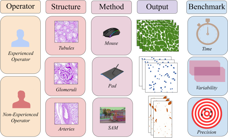
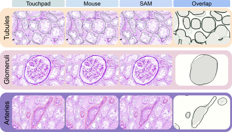
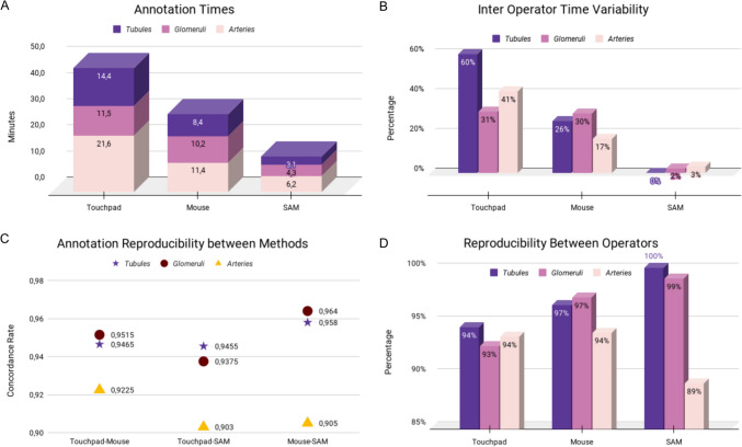
The development of reliable artificial intelligence (AI) algorithms in pathology often depends on ground truth provided by annotation of whole slide images (WSI), a time-consuming and operator-dependent process. A comparative analysis of different annotation approaches is performed to streamline this process. Two pathologists annotated renal tissue using semi-automated (Segment Anything Model, SAM)) and manual devices (touchpad vs mouse). A comparison was conducted in terms of working time, reproducibility (overlap fraction), and precision (0 to 10 accuracy rated by two expert nephropathologists) among different methods and operators. The impact of different displays on mouse performance was evaluated. Annotations focused on three tissue compartments: tubules (57 annotations), glomeruli (53 annotations), and arteries (58 annotations). The semi-automatic approach was the fastest and had the least inter-observer variability, averaging 13.6 ± 0.2 min with a difference (Δ) of 2%, followed by the mouse (29.9 ± 10.2, Δ = 24%), and the touchpad (47.5 ± 19.6 min, Δ = 45%). The highest reproducibility in tubules and glomeruli was achieved with SAM (overlap values of 1 and 0.99 compared to 0.97 for the mouse and 0.94 and 0.93 for the touchpad), though SAM had lower reproducibility in arteries (overlap value of 0.89 compared to 0.94 for both the mouse and touchpad). No precision differences were observed between operators (p = 0.59). Using non-medical monitors increased annotation times by 6.1%. The future employment of semi-automated and AI-assisted approaches can significantly speed up the annotation process, improving the ground truth for AI tool development.

摘要

病理学中可靠人工智能(AI)算法的开发通常依赖于通过全切片图像(WSI)标注提供的真实数据,这是一个耗时且依赖操作人员的过程。为简化这一过程,对不同的标注方法进行了对比分析。两名病理学家使用半自动设备(分割一切模型,即SAM)和手动设备(触摸板与鼠标)对肾组织进行标注。在不同方法和操作人员之间,就工作时间、可重复性(重叠率)和精确度(由两名肾脏病理专家给出0至10的准确度评分)进行了比较。评估了不同显示器对鼠标操作性能的影响。标注集中在三个组织区域:肾小管(57个标注)、肾小球(53个标注)和动脉(58个标注)。半自动方法最快,观察者间变异性最小,平均用时13.6±0.2分钟,差异(Δ)为2%,其次是鼠标(29.9±10.2分钟,Δ=24%)和触摸板(47.5±19.6分钟,Δ=45%)。SAM在肾小管和肾小球方面实现了最高的可重复性(重叠值分别为1和0.99,而鼠标为0.97,触摸板为0.94和0.93),不过SAM在动脉方面的可重复性较低(重叠值为0.89,而鼠标和触摸板均为0.94)。未观察到操作人员之间在精确度上存在差异(p=0.59)。使用非医用显示器会使标注时间增加6.1%。未来采用半自动和AI辅助方法可显著加快标注过程,为AI工具开发提供更好的真实数据。

https://cdn.ncbi.nlm.nih.gov/pmc/blobs/5aa7/11950598/9d5944812328/10278_2024_1248_Fig4_HTML.jpg
https://cdn.ncbi.nlm.nih.gov/pmc/blobs/5aa7/11950598/2fb6abb8cd58/10278_2024_1248_Fig1_HTML.jpg
https://cdn.ncbi.nlm.nih.gov/pmc/blobs/5aa7/11950598/464b08851f50/10278_2024_1248_Fig2_HTML.jpg
https://cdn.ncbi.nlm.nih.gov/pmc/blobs/5aa7/11950598/6f689d8735c6/10278_2024_1248_Fig3_HTML.jpg
https://cdn.ncbi.nlm.nih.gov/pmc/blobs/5aa7/11950598/9d5944812328/10278_2024_1248_Fig4_HTML.jpg

相似文献

1
Improving the Annotation Process in Computational Pathology: A Pilot Study with Manual and Semi-automated Approaches on Consumer and Medical Grade Devices.改进计算病理学中的标注过程:一项关于消费级和医疗级设备的手动及半自动方法的试点研究。
J Imaging Inform Med. 2025 Apr;38(2):1112-1119. doi: 10.1007/s10278-024-01248-x. Epub 2024 Sep 4.
2
Overcoming an Annotation Hurdle: Digitizing Pen Annotations from Whole Slide Images.跨越注释障碍:将来自全切片图像的手写注释数字化
J Pathol Inform. 2021 Feb 23;12:9. doi: 10.4103/jpi.jpi_85_20. eCollection 2021.
3
Annotation Practices in Computational Pathology: A European Society of Digital and Integrative Pathology (ESDIP) Survey Study.计算病理学中的注释实践:欧洲数字与整合病理学学会(ESDIP)调查研究
Lab Invest. 2025 Mar;105(3):102203. doi: 10.1016/j.labinv.2024.102203. Epub 2024 Nov 29.
4
Semantic annotation for computational pathology: multidisciplinary experience and best practice recommendations.计算病理学的语义标注:多学科经验和最佳实践建议。
J Pathol Clin Res. 2022 Mar;8(2):116-128. doi: 10.1002/cjp2.256. Epub 2022 Jan 10.
5
A comparative study of the inter-observer variability on Gleason grading against Deep Learning-based approaches for prostate cancer.基于深度学习的前列腺癌 Gleason 分级方法的观察者间变异性比较研究。
Comput Biol Med. 2023 Jun;159:106856. doi: 10.1016/j.compbiomed.2023.106856. Epub 2023 Apr 6.
6
Development and Validation of an Artificial Intelligence-Powered Platform for Prostate Cancer Grading and Quantification.开发和验证一个用于前列腺癌分级和定量的人工智能平台。
JAMA Netw Open. 2021 Nov 1;4(11):e2132554. doi: 10.1001/jamanetworkopen.2021.32554.
7
Combined colour deconvolution and artificial intelligence approach for region-selective immunohistochemical labelling quantification: The example of alpha smooth muscle actin in mouse kidney.联合色彩分解和人工智能方法进行区域选择性免疫组织化学标记定量:以小鼠肾脏中的α平滑肌肌动蛋白为例。
J Biophotonics. 2024 Jan;17(1):e202300244. doi: 10.1002/jbio.202300244. Epub 2023 Nov 7.
8
Leverage Weakly Annotation to Pixel-wise Annotation via Zero-shot Segment Anything Model for Molecular-empowered Learning.通过零样本分割一切模型利用弱注释进行逐像素注释以实现分子增强学习
Proc SPIE Int Soc Opt Eng. 2024 Feb;12933. doi: 10.1117/12.3006577. Epub 2024 Apr 3.
9
Semi-automated renal region of interest selection method using the double-threshold technique: inter-operator variability in quantitating 99mTc-MAG3 renal uptake.使用双阈值技术的半自动肾脏感兴趣区选择方法:99mTc-MAG3肾脏摄取定量中的操作者间变异性
Eur J Nucl Med. 1998 Jan;25(1):55-9.
10
Quantitative assessment of myocardial fibrosis by digital image analysis: An adjunctive tool for pathologist "ground truth".通过数字图像分析对心肌纤维化进行定量评估:病理学家“真实数据”的辅助工具。
Cardiovasc Pathol. 2023 Jul-Aug;65:107541. doi: 10.1016/j.carpath.2023.107541. Epub 2023 Apr 29.

引用本文的文献

1
Automating liver biopsy segmentation with a robust, open-source tool for pathology research: the HOTSPoT model.利用一个用于病理学研究的强大开源工具实现肝活检分割自动化:HOTSPoT模型。
NPJ Digit Med. 2025 Jul 18;8(1):455. doi: 10.1038/s41746-025-01870-1.
2
Analyzing the performance of biomedical time-series segmentation with electrophysiology data.利用电生理数据分析生物医学时间序列分割的性能。
Sci Rep. 2025 Apr 6;15(1):11776. doi: 10.1038/s41598-025-90533-y.

本文引用的文献

1
Sequential Patching Lattice for Image Classification and Enquiry: Streamlining Digital Pathology Image Processing.序列补丁晶格在图像分类和查询中的应用:简化数字病理学图像处理。
Am J Pathol. 2024 Oct;194(10):1898-1912. doi: 10.1016/j.ajpath.2024.06.007. Epub 2024 Jul 18.
2
Benchmarking digital displays (monitors) for histological diagnoses: the nephropathology use case.用于组织学诊断的数字显示器(监视器)基准测试:肾病理学用例。
J Clin Pathol. 2024 Nov 21. doi: 10.1136/jcp-2024-209418.
3
Perspective of a Pathologist on Benchmark Strategies for Artificial Intelligence Development in Organ Transplantation.
病理学家视角下的器官移植人工智能发展基准策略。
Crit Rev Oncog. 2023;28(3):1-6. doi: 10.1615/CritRevOncog.2023048797.
4
Segment Anything by Meta as a foundation model for image segmentation: a new era for histopathological images.Meta公司的Segment Anything作为图像分割的基础模型:开启组织病理学图像的新时代。
Pathology. 2023 Dec;55(7):1017-1020. doi: 10.1016/j.pathol.2023.09.003. Epub 2023 Sep 26.
5
Time for a full digital approach in nephropathology: a systematic review of current artificial intelligence applications and future directions.肾脏病学全面数字化时代:当前人工智能应用及未来方向的系统综述。
J Nephrol. 2024 Jan;37(1):65-76. doi: 10.1007/s40620-023-01775-w. Epub 2023 Sep 28.
6
Performance and limitations of a supervised deep learning approach for the histopathological Oxford Classification of glomeruli with IgA nephropathy.用于IgA肾病肾小球组织病理学牛津分类的监督式深度学习方法的性能与局限性
Comput Methods Programs Biomed. 2023 Dec;242:107814. doi: 10.1016/j.cmpb.2023.107814. Epub 2023 Sep 13.
7
Segment anything model for medical image analysis: An experimental study.用于医学图像分析的分割模型:一项实验研究。
Med Image Anal. 2023 Oct;89:102918. doi: 10.1016/j.media.2023.102918. Epub 2023 Aug 2.
8
Standardized Clinical Annotation of Digital Histopathology Slides at the Point of Diagnosis.在诊断点对数字组织病理学幻灯片进行标准化临床注释。
Mod Pathol. 2023 Nov;36(11):100297. doi: 10.1016/j.modpat.2023.100297. Epub 2023 Aug 4.
9
The slow-paced digital evolution of pathology: lights and shadows from a multifaceted board.病理学的数字化演进缓慢:多方面董事会的光明与阴影。
Pathologica. 2023 Jun;115(3):127-136. doi: 10.32074/1591-951X-868.
10
Interoperable slide microscopy viewer and annotation tool for imaging data science and computational pathology.可互操作的幻灯片显微镜查看器和注释工具,用于成像数据科学和计算病理学。
Nat Commun. 2023 Mar 22;14(1):1572. doi: 10.1038/s41467-023-37224-2.