Verdi Serena, Rutherford Saige, Fraza Charlotte, Tosun Duygu, Altmann Andre, Raket Lars Lau, Schott Jonathan M, Marquand Andre F, Cole James H
Centre for Medical Image Computing, University College London, London, UK.
Dementia Research Centre, UCL Queen Square Institute of Neurology, London, UK.
Alzheimers Dement. 2024 Oct;20(10):6998-7012. doi: 10.1002/alz.14174. Epub 2024 Sep 5.
Neuroanatomical normative modeling captures individual variability in Alzheimer's disease (AD). Here we used normative modeling to track individuals' disease progression in people with mild cognitive impairment (MCI) and patients with AD.
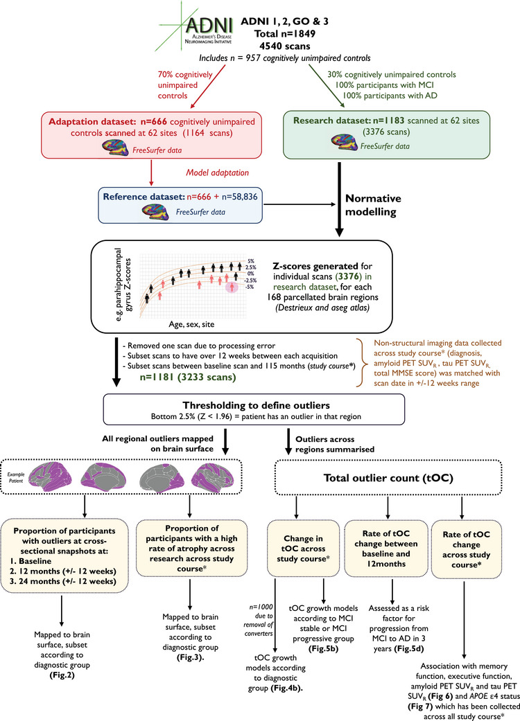
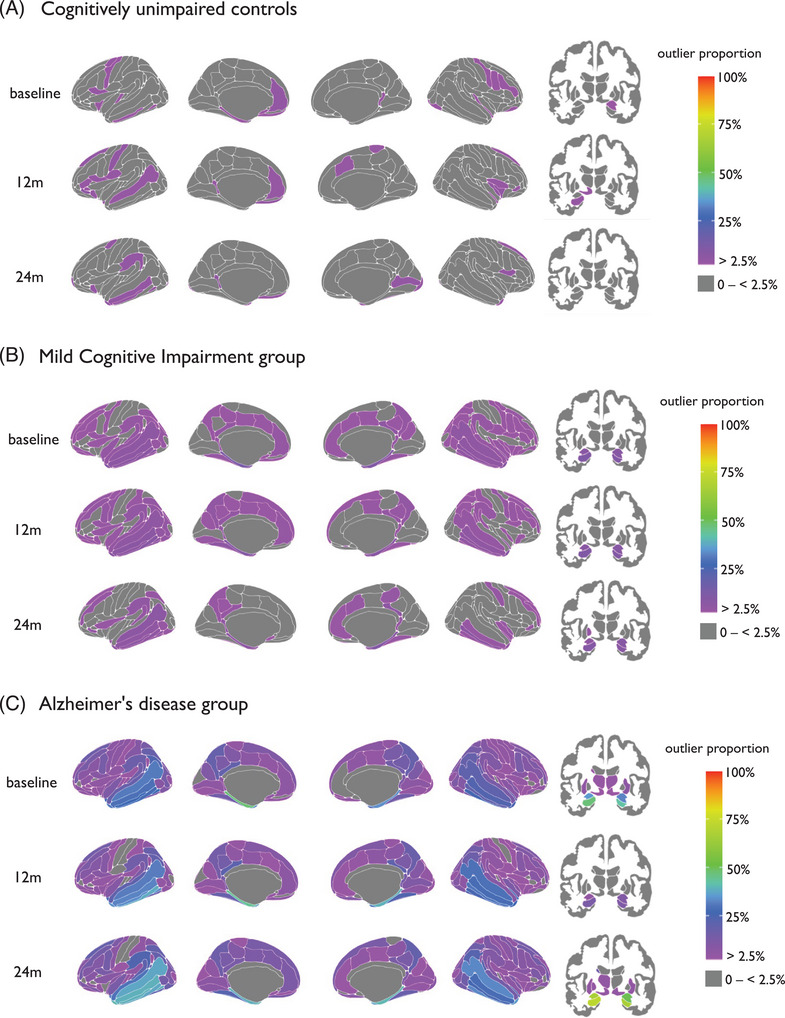
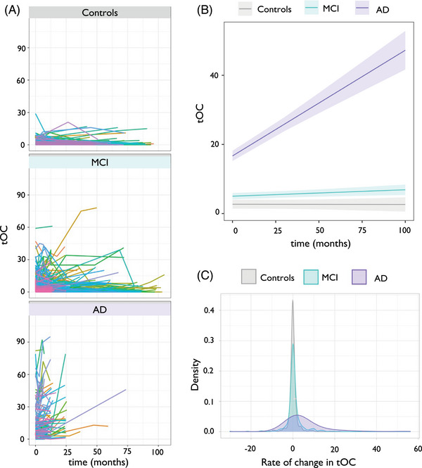
Cortical and subcortical normative models were generated using healthy controls (n ≈ 58k). These models were used to calculate regional z scores in 3233 T1-weighted magnetic resonance imaging time-series scans from 1181 participants. Regions with z scores < -1.96 were classified as outliers mapped on the brain and summarized by total outlier count (tOC).
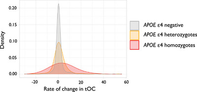
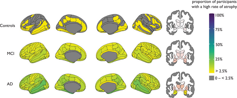
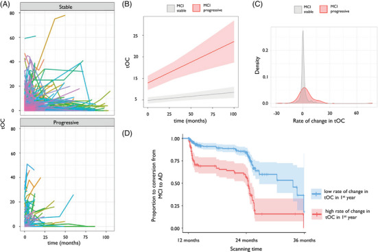
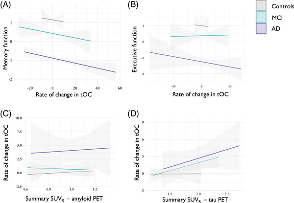
tOC increased in AD and in people with MCI who converted to AD and also correlated with multiple non-imaging markers. Moreover, a higher annual rate of change in tOC increased the risk of progression from MCI to AD. Brain outlier maps identified the hippocampus as having the highest rate of change.
Individual patients' atrophy rates can be tracked by using regional outlier maps and tOC.
Neuroanatomical normative modeling was applied to serial Alzheimer's disease (AD) magnetic resonance imaging (MRI) data for the first time. Deviation from the norm (outliers) of cortical thickness or brain volume was computed in 3233 scans. The number of brain-structure outliers increased over time in people with AD. Patterns of change in outliers varied markedly between individual patients with AD. People with mild cognitive impairment whose outliers increased over time had a higher risk of progression from AD.
神经解剖学规范模型捕捉了阿尔茨海默病(AD)中的个体变异性。在此,我们使用规范模型来追踪轻度认知障碍(MCI)患者和AD患者的疾病进展情况。
利用健康对照者(n≈58000)生成皮质和皮质下规范模型。这些模型用于计算来自1181名参与者的3233次T1加权磁共振成像时间序列扫描中的区域z分数。z分数< -1.96的区域被分类为大脑上映射的异常值,并通过总异常值计数(tOC)进行汇总。
AD患者以及转化为AD的MCI患者的tOC增加,并且与多个非影像学标志物相关。此外,tOC的较高年变化率增加了从MCI进展为AD的风险。脑异常值图显示海马体的变化率最高。
可以使用区域异常值图和tOC来追踪个体患者的萎缩率。
神经解剖学规范模型首次应用于阿尔茨海默病(AD)的系列磁共振成像(MRI)数据。在3233次扫描中计算了皮质厚度或脑容量与规范的偏差(异常值)。AD患者的脑结构异常值数量随时间增加。AD个体患者之间异常值的变化模式明显不同。随着时间推移异常值增加的轻度认知障碍患者进展为AD的风险更高。