Fritsch Hanna, Tumeltshammer Rebecca, Hachenberg Jens, Warm Mathias, Krug Barbara, Malter Wolfram, Eichler Christian
Breast Cancer Center, St. Franziskus-Hospital, Münster, Germany.
Department of Gynecology and Obstetrics, Faculty of Medicine, University of Cologne, Cologne, Germany.
Cancer Diagn Progn. 2024 Sep 1;4(5):599-604. doi: 10.21873/cdp.10369. eCollection 2024 Sep-Oct.
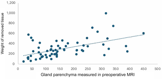
BACKGROUND/AIM: This study examined the influence of preoperative MRI on the choice of implant volume in patients undergoing subcutaneous mastectomy with immediate breast reconstruction. It was postulated that preoperative MRI scans can adequately estimate glandular tissue, which in turn correlates with implant size.
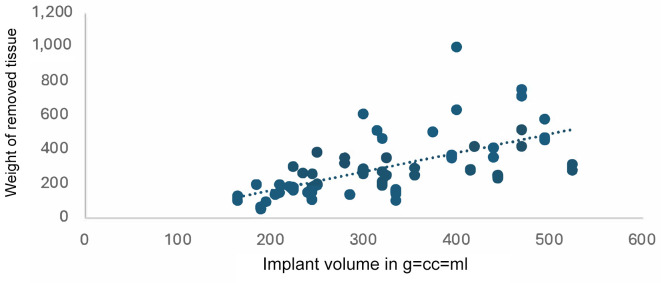
Preoperative and postoperative MRI scans were used in oncological and prophylactical subcutaneous mastectomy scenarios in 67 cases at the Department of Gynaecology, Breast Cancer Center, University of Cologne, Germany. The preoperative MRI was used to estimate the resected tissue and the postoperative MRI was used to scan for residual glandular tissue. In addition, a correlation found by Malter et al. in 2021 was evaluated with the available data.
Preoperative MRIs result in an adequate estimation of resected tissue. This in turn correlates with implant volume. The correlation by Malter et al. also holds when estimating implant volume. The likelihood of residual gland was low if the preoperatively estimate volume was removed.
Our results indicate that the use of preoperative and postoperative MRI scans for subcutaneous mastectomies is advantageous. We suggest a routine estimation of glandular tissue, especially for small breasts.
背景/目的:本研究探讨了术前MRI对接受皮下乳房切除术并即刻乳房重建患者植入体体积选择的影响。研究假设术前MRI扫描能够充分估计腺体组织,而腺体组织又与植入体大小相关。
德国科隆大学乳腺癌中心妇科对67例患者在肿瘤性和预防性皮下乳房切除术中使用了术前和术后MRI扫描。术前MRI用于估计切除组织,术后MRI用于扫描残留腺体组织。此外,利用现有数据评估了Malter等人在2021年发现的相关性。
术前MRI能够充分估计切除组织。这进而与植入体体积相关。在估计植入体体积时,Malter等人发现的相关性同样成立。如果术前估计的体积被切除,残留腺体的可能性较低。
我们的结果表明,术前和术后MRI扫描用于皮下乳房切除术具有优势。我们建议常规估计腺体组织,尤其是对于小乳房。