de Sena Bruna Voltolin, Turquete Paula Baêta da Silva Rios, Pimentel Pedro Antônio Bronhara, Almeida Isabella Oliveira, Lavalle Gleidice Eunice, Nakagaki Karen Yumi Ribeiro, Giuliano Antonio, Paes Paulo Ricardo de Oliveira, Horta Rodrigo Dos Santos
Department of Veterinary Medicine and Surgery, Veterinary School, Universidade Federal de Minas Gerais, Belo Horizonte, Minas Gerais, Brazil.
Director and Technical Responsible at CELULAVET, Belo Horizonte, Brazil.
Front Vet Sci. 2024 Aug 22;11:1397592. doi: 10.3389/fvets.2024.1397592. eCollection 2024.
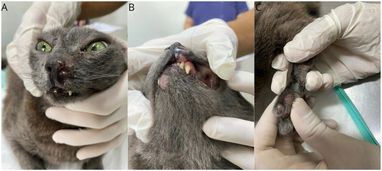
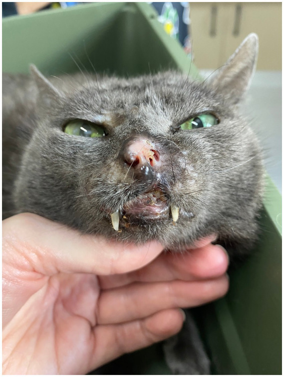
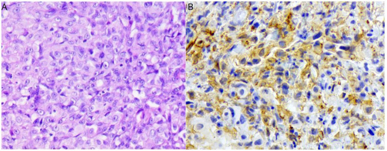
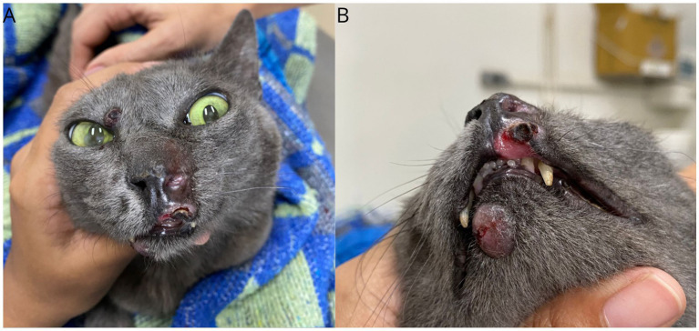
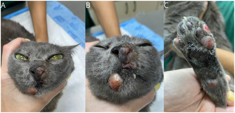
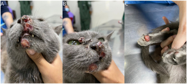
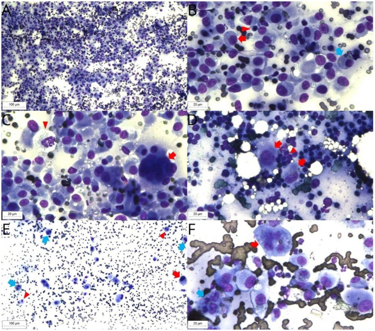
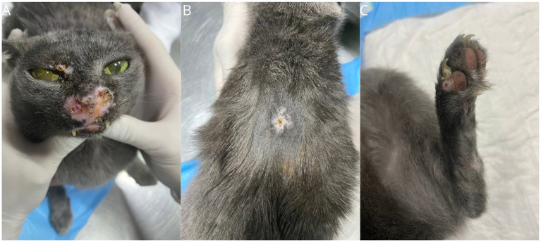
Feline histiocytic diseases are uncommon and rarely reported. Feline progressive histiocytosis (FPH) is the most common histiocytic disease in cats, predominantly affecting middle-aged animals. The most common presentation is the cutaneous form with solitary or multiple cutaneous nodules. A female, mixed-breed 6-year-old cat was presented with a 9-month history of a nodule in the nasal planum and was diagnosed by histopathology with histiocytic proliferation. At the time of diagnosis, new nodules were discovered on the lower lip, digit, and two lesions in the tail region, with the largest measuring 1.5 cm. Supplementary immunohistochemistry, showed immunolabeling for Iba-1 that in combination with the clinical course of the disease, confirmed the diagnosis of FPH. No response to chemotherapy treatment with lomustine alternated with doxorubicin was achieved. Toceranib phosphate resulted in a transient response and, stable disease for a short period (6 weeks). Electrochemotherapy with bleomycin was initiated and resulted in partial remission. Later on, chlorambucil was also started. Ultimately, the combination of all three treatments led to a complete response and disappearance of all the lesions. FPH is considered a disease resistant to various treatments, and effective treatments have not been reported. In this case report, we describe a successful multimodal therapeutic approach that resulted in complete resolution of the FPH and long-term survival (460 days without external lesions at the time of death). Further studies are necessary to confirm the efficacy of this therapeutic approach.
猫组织细胞增多症并不常见,报道也很少。猫进行性组织细胞增多症(FPH)是猫最常见的组织细胞疾病,主要影响中年动物。最常见的表现形式是皮肤型,表现为单个或多个皮肤结节。一只6岁的雌性混种猫因鼻平面出现一个结节已有9个月病史前来就诊,经组织病理学诊断为组织细胞增殖。在诊断时,在下唇、趾部以及尾部区域发现了新的结节,最大的结节直径为1.5厘米。补充免疫组化显示Iba-1免疫标记阳性,结合疾病的临床病程,确诊为FPH。使用洛莫司汀交替多柔比星进行化疗未取得疗效。磷酸托西替尼产生了短暂反应,并使病情在短时间内(6周)保持稳定。开始使用博来霉素进行电化学疗法,结果病情部分缓解。后来也开始使用苯丁酸氮芥。最终,三种治疗方法联合使用导致完全缓解,所有病变消失。FPH被认为是一种对各种治疗均有抵抗性的疾病,尚未有有效治疗方法的报道。在本病例报告中,我们描述了一种成功的多模式治疗方法,该方法使FPH完全消退,并实现了长期存活(死亡时无外部病变存活460天)。需要进一步研究以证实这种治疗方法的疗效。