• 文献检索
  • 文档翻译
  • 深度研究
  • 学术资讯
  • Suppr Zotero 插件Zotero 插件
  • 邀请有礼
  • 套餐&价格
  • 历史记录
应用&插件
Suppr Zotero 插件Zotero 插件浏览器插件Mac 客户端Windows 客户端微信小程序
定价
高级版会员购买积分包购买API积分包
服务
文献检索文档翻译深度研究API 文档MCP 服务
关于我们
关于 Suppr公司介绍联系我们用户协议隐私条款
关注我们

Suppr 超能文献

核心技术专利:CN118964589B侵权必究
粤ICP备2023148730 号-1Suppr @ 2026

文献检索

告别复杂PubMed语法,用中文像聊天一样搜索,搜遍4000万医学文献。AI智能推荐,让科研检索更轻松。

立即免费搜索

文件翻译

保留排版,准确专业,支持PDF/Word/PPT等文件格式,支持 12+语言互译。

免费翻译文档

深度研究

AI帮你快速写综述,25分钟生成高质量综述,智能提取关键信息,辅助科研写作。

立即免费体验

腰背支的高保真三维建模

High-fidelity 3D modelling of the lumbar dorsal rami.

作者信息

Tran John, Campisi Emma, Roa Agudelo Alexandria, Agur Anne Mr, Loh Eldon

机构信息

Department of Physical Medicine and Rehabilitation, Parkwood Institute, London, Ontario, N6C 0A7, Canada.

Lawson Health Research Institute, London, Ontario, N6C 2R5, Canada.

出版信息

Interv Pain Med. 2024 Mar 7;3(1):100401. doi: 10.1016/j.inpm.2024.100401. eCollection 2024 Mar.

DOI:10.1016/j.inpm.2024.100401
PMID:39239499
原文链接:https://pmc.ncbi.nlm.nih.gov/articles/PMC11373041/
Abstract

OBJECTIVE

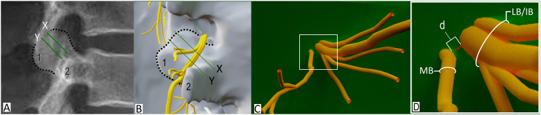
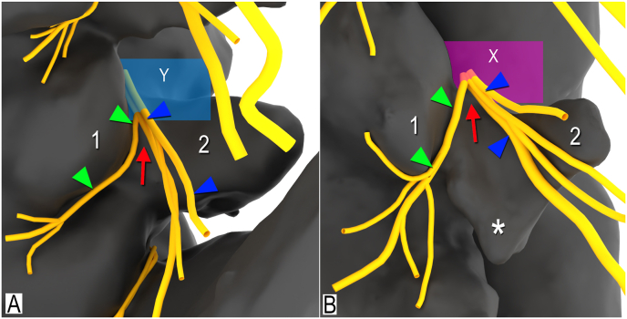
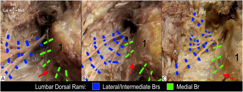
Lumbar medial branch denervation is commonly used to treat chronic facetogenic low back pain. Controversy exists regarding risk to adjacent neural structures. The objectives of this cadaveric study were to: (1) dissect, digitize, and model in 3D the branches of the first (L1) to fifth (L5) lumbar dorsal rami located near the junction of the transverse process and lateral neck of the superior articular process; and (2) quantify the minimal distance between the lateral/intermediate and medial branches at the anterior quarter and midpoint of the lateral neck of the superior articular process.

DESIGN

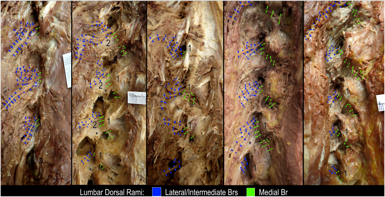
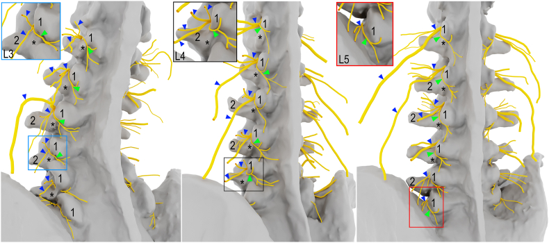
Eighteen formalin-embalmed specimens were dissected, digitized and modeled in 3D. The high-fidelity 3D models were used to compare branching patterns and quantify the mean minimal distance between the lateral/intermediate and medial branches of the lumbar dorsal ramus at the anterior quarter and midpoint of the lateral neck of the superior articular process. A Two-way ANOVA was performed to determine if difference of mean distances was significant.

RESULTS

There was variability in the branching pattern of the lumbar dorsal rami. In 46 cases (51.1%) the lumbar dorsal ramus divided into 2 branches, in 41 cases (45.6%) into 3, and in 3 cases (3.3%) 4. The mean minimal distance between the lateral/intermediate and medial branches was significantly greater at the midpoint (3.2 ± 2.5 mm) than the anterior quarter (1.2 ± 1.8 mm) of the lateral neck of superior articular process.

CONCLUSION

Minimal distance measurements between the branches of the lumbar dorsal rami at the anterior quarter and midpoint of the lateral neck of the superior articular process were computed. When placing the distal end of the needle tip at the anterior quarter of the lateral neck of the superior articular process, the smaller mean minimal distance between the branches suggests there is a greater risk for inadvertent denervation of the lateral/intermediate branches. Further anatomical and clinical investigations are required.

摘要

目的

腰椎内侧支去神经术常用于治疗慢性小关节源性下腰痛。关于其对相邻神经结构的风险存在争议。本尸体研究的目的是:(1)对位于横突与上关节突外侧颈交界处附近的第一(L1)至第五(L5)腰背支进行解剖、数字化并建立三维模型;(2)量化上关节突外侧颈前四分之一处及中点处外侧/中间支与内侧支之间的最小距离。

设计

对18个用福尔马林防腐的标本进行解剖、数字化并建立三维模型。使用高保真三维模型比较分支模式,并量化上关节突外侧颈前四分之一处及中点处腰背支外侧/中间支与内侧支之间的平均最小距离。进行双向方差分析以确定平均距离差异是否显著。

结果

腰背支的分支模式存在变异性。在46例(51.1%)中,腰背支分为2支,41例(45.6%)分为3支,3例(3.3%)分为4支。上关节突外侧颈中点处外侧/中间支与内侧支之间的平均最小距离(3.2±2.5毫米)显著大于外侧颈前四分之一处(1.2±1.8毫米)。

结论

计算了上关节突外侧颈前四分之一处及中点处腰背支各分支之间的最小距离。当将针尖远端置于上关节突外侧颈前四分之一处时,各分支之间较小的平均最小距离表明外侧/中间支意外去神经的风险更大。需要进一步的解剖学和临床研究。

https://cdn.ncbi.nlm.nih.gov/pmc/blobs/8448/11373041/28fdc63e4c09/gr5.jpg
https://cdn.ncbi.nlm.nih.gov/pmc/blobs/8448/11373041/2d735c8bdb70/gr1.jpg
https://cdn.ncbi.nlm.nih.gov/pmc/blobs/8448/11373041/421915118d56/gr2.jpg
https://cdn.ncbi.nlm.nih.gov/pmc/blobs/8448/11373041/c00c2a7293d3/gr3.jpg
https://cdn.ncbi.nlm.nih.gov/pmc/blobs/8448/11373041/d7ddc8546b4a/gr4.jpg
https://cdn.ncbi.nlm.nih.gov/pmc/blobs/8448/11373041/28fdc63e4c09/gr5.jpg

相似文献

1
High-fidelity 3D modelling of the lumbar dorsal rami.腰背支的高保真三维建模
Interv Pain Med. 2024 Mar 7;3(1):100401. doi: 10.1016/j.inpm.2024.100401. eCollection 2024 Mar.
2
3D nerve proximity mapping of the medial branch of lumbar dorsal ramus: An anatomical study.腰背侧支内侧支的三维神经毗邻关系图谱:一项解剖学研究。
Interv Pain Med. 2024 May 15;3(2):100414. doi: 10.1016/j.inpm.2024.100414. eCollection 2024 Jun.
3
Anatomical study of the medial branches of the lumbar dorsal rami: implications for image-guided intervention.腰背部神经后支内侧支的解剖学研究:对影像引导介入的意义
Reg Anesth Pain Med. 2022 May 19. doi: 10.1136/rapm-2022-103653.
4
Endoscopically guided foraminal and dorsal rhizotomy for chronic axial back pain based on cadaver and endoscopically visualized anatomic study.基于尸体和内镜可视化解剖学研究的内镜引导下椎间孔和背根神经切断术治疗慢性轴性背痛
Int J Spine Surg. 2014 Dec 1;8. doi: 10.14444/1023. eCollection 2014.
5
The anatomy of the so-called "articular nerves" and their relationship to facet denervation in the treatment of low-back pain.所谓“关节神经”的解剖结构及其在腰痛治疗中与小关节去神经支配的关系。
J Neurosurg. 1979 Aug;51(2):172-7. doi: 10.3171/jns.1979.51.2.0172.
6
Anatomical Observation and Clinical Significance of the Medial Branch of the Lumbar Dorsal Rami.腰椎背侧支内侧支的解剖观察及临床意义。
Spine (Phila Pa 1976). 2024 Jun 1;49(11):E164-E172. doi: 10.1097/BRS.0000000000004975. Epub 2024 Feb 29.
7
Investigation of the anatomical course of articular branches to the thoracic zygapophysial joints.对胸椎间关节的关节支解剖走行的研究。
Interv Pain Med. 2024 Mar 11;3(1):100399. doi: 10.1016/j.inpm.2024.100399. eCollection 2024 Mar.
8
Anatomic-Topographic Investigation of the Branches of the Dorsal Ramus of Thoracic Spinal Nerves.胸神经背支分支的解剖-拓扑学研究。
Pain Med. 2022 Oct 29;23(11):1869-1874. doi: 10.1093/pm/pnac072.
9
Anatomical analysis of medial branches of dorsal rami of cervical nerves for radiofrequency thermocoagulation.颈椎神经背支内侧支射频热凝的解剖学分析。
Reg Anesth Pain Med. 2014 Nov-Dec;39(6):465-71. doi: 10.1097/AAP.0000000000000175.
10
[Anatomical background of low back pain: variability and degeneration of the lumbar spinal canal and intervertebral disc].[腰痛的解剖学背景:腰椎管和椎间盘的变异性与退变]
Schmerz. 2001 Dec;15(6):418-24. doi: 10.1007/s004820100026.

引用本文的文献

1
Lumbar facet joint denervation targeting the medial branch in the sub-mammillary fossa: An anatomical optimization study.针对乳突下窝内侧支的腰椎小关节去神经支配:一项解剖学优化研究。
Interv Pain Med. 2025 Apr 22;4(2):100586. doi: 10.1016/j.inpm.2025.100586. eCollection 2025 Jun.
2
Targeting the lateral neck of superior articular process and sub-mammillary fossa for lumbar medial branch radiofrequency ablation: A case series.靶向腰椎内侧支射频消融术中的上关节突外侧颈部和乳突下窝:病例系列
Interv Pain Med. 2024 Nov 30;3(4):100533. doi: 10.1016/j.inpm.2024.100533. eCollection 2024 Dec.
3
Evaluation of a novel nerve ablation technique to relieve lower back pain: a cadaveric feasibility pilot study.

本文引用的文献

1
Parasagittal needle placement approach for lumbar medial branch denervation: a brief technical report.经椎间孔腰椎内侧支射频消融术的矢状位进针方法:简要技术报告。
Reg Anesth Pain Med. 2024 Aug 5;49(8):609-614. doi: 10.1136/rapm-2023-105152.
2
Abnormal Paresthesias Associated With Radiofrequency Ablation of Lumbar Medial Branch Nerves: A Case Report.与腰椎内侧支神经射频消融相关的异常感觉异常:一例报告
Cureus. 2023 Feb 19;15(2):e35176. doi: 10.7759/cureus.35176. eCollection 2023 Feb.
3
Reply to letter to editor regarding 'anatomical study of the medial branches of the lumbar dorsal rami: implications for image guided intervention'.
一种新型神经消融技术缓解下背部疼痛的评估:一项尸体可行性初步研究。
Pain Med. 2025 Feb 1;26(2):70-75. doi: 10.1093/pm/pnae112.
4
Optimal caudal needle angulation for lumbar medial branch denervation: A 3D cadaveric and clinical imaging comparison study.腰椎内侧支去神经术的最佳尾侧进针角度:一项三维尸体与临床影像对比研究。
Interv Pain Med. 2024 Aug 19;3(3):100433. doi: 10.1016/j.inpm.2024.100433. eCollection 2024 Sep.
对致编辑的信的回复,信的主题为“腰背部神经后支内侧支的解剖学研究:对影像引导介入的意义”
Reg Anesth Pain Med. 2023 Feb;48(2):95-96. doi: 10.1136/rapm-2022-104168. Epub 2022 Nov 16.
4
Letter to the editor regarding 'Anatomical study of the medial branches of the lumbar dorsal rami: implications for image-guided intervention'.致编辑的信:关于“腰背部神经后支内侧支的解剖学研究:对影像引导介入的意义”
Reg Anesth Pain Med. 2023 Feb;48(2):94. doi: 10.1136/rapm-2022-104119. Epub 2022 Nov 16.
5
Quantification of Needle Angles for Traditional Lumbar Medial Branch Radiofrequency Ablation: An Osteological Study.传统腰椎内侧支射频消融术进针角度的量化:一项骨骼学研究。
Pain Med. 2023 May 2;24(5):488-495. doi: 10.1093/pm/pnac160.
6
Prospective Within Subject Comparison of Fluoroscopically Guided Lumbosacral Facet Joint Radiofrequency Ablation Using a Multi-Tined (Trident) Versus Conventional Monopolar Cannula.前瞻性自身对照研究:多齿(三叉)与传统单极套管经皮射频消融腰椎关节突关节。
Pain Physician. 2022 Aug;25(5):391-399.
7
Anatomical study of the medial branches of the lumbar dorsal rami: implications for image-guided intervention.腰背部神经后支内侧支的解剖学研究:对影像引导介入的意义
Reg Anesth Pain Med. 2022 May 19. doi: 10.1136/rapm-2022-103653.
8
Update of Utilization Patterns of Facet Joint Interventions in Managing Spinal Pain from 2000 to 2018 in the US Fee-for-Service Medicare Population.2000 年至 2018 年美国按服务收费的医疗保险人群中,治疗脊柱疼痛的关节突关节介入治疗利用模式的更新。
Pain Physician. 2020 Mar;23(2):E133-E149.
9
Systematic Review of the Effectiveness of Lumbar Medial Branch Thermal Radiofrequency Neurotomy, Stratified for Diagnostic Methods and Procedural Technique.腰椎内侧支热射频神经切断术有效性的系统评价,按诊断方法和手术技术分层
Pain Med. 2020 Jun 1;21(6):1122-1141. doi: 10.1093/pm/pnz349.
10
Anatomical etiology of "pseudo-sciatica" from superior cluneal nerve entrapment: a laboratory investigation.臀上皮神经卡压所致“假性坐骨神经痛”的解剖学病因:一项实验室研究
J Pain Res. 2017 Nov 1;10:2539-2545. doi: 10.2147/JPR.S142115. eCollection 2017.