• 文献检索
  • 文档翻译
  • 深度研究
  • 学术资讯
  • Suppr Zotero 插件Zotero 插件
  • 邀请有礼
  • 套餐&价格
  • 历史记录
应用&插件
Suppr Zotero 插件Zotero 插件浏览器插件Mac 客户端Windows 客户端微信小程序
定价
高级版会员购买积分包购买API积分包
服务
文献检索文档翻译深度研究API 文档MCP 服务
关于我们
关于 Suppr公司介绍联系我们用户协议隐私条款
关注我们

Suppr 超能文献

核心技术专利:CN118964589B侵权必究
粤ICP备2023148730 号-1Suppr @ 2026

文献检索

告别复杂PubMed语法,用中文像聊天一样搜索,搜遍4000万医学文献。AI智能推荐,让科研检索更轻松。

立即免费搜索

文件翻译

保留排版,准确专业,支持PDF/Word/PPT等文件格式,支持 12+语言互译。

免费翻译文档

深度研究

AI帮你快速写综述,25分钟生成高质量综述,智能提取关键信息,辅助科研写作。

立即免费体验

β-arrestin-2 通过磷脂酰肌醇 4,5-二磷酸的预激活的分子机制。

Molecular mechanism of β-arrestin-2 pre-activation by phosphatidylinositol 4,5-bisphosphate.

机构信息

School of Pharmacy, Sungkyunkwan University, 2066 Seobu-ro, Jangan-gu, Suwon, 16419, Republic of Korea.

出版信息

EMBO Rep. 2024 Oct;25(10):4190-4205. doi: 10.1038/s44319-024-00239-x. Epub 2024 Sep 6.

DOI:10.1038/s44319-024-00239-x
PMID:39242774
原文链接:https://pmc.ncbi.nlm.nih.gov/articles/PMC11467438/
Abstract

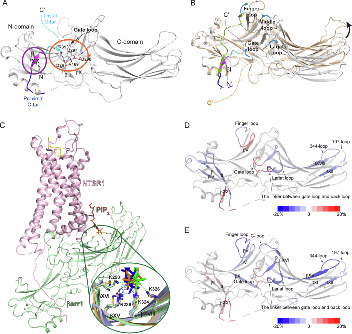
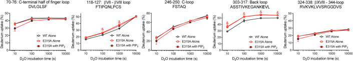
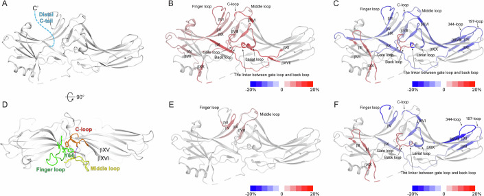
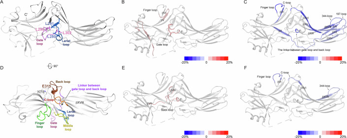
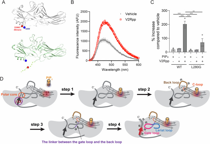
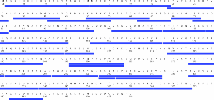
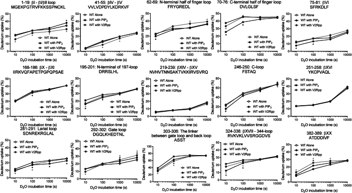
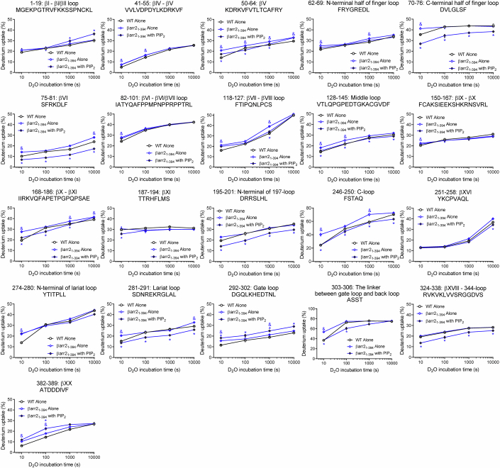
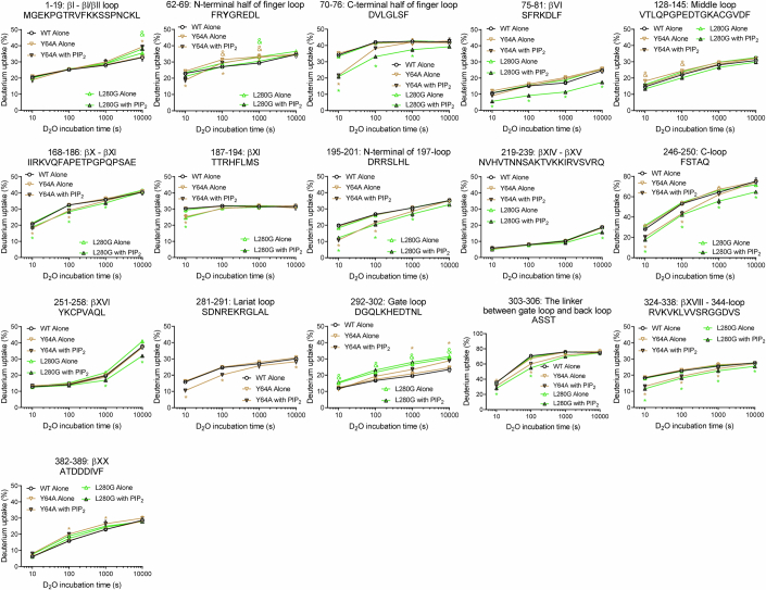
Phosphorylated residues of G protein-coupled receptors bind to the N-domain of arrestin, resulting in the release of its C-terminus. This induces further allosteric conformational changes, such as polar core disruption, alteration of interdomain loops, and domain rotation, which transform arrestins into the receptor-activated state. It is widely accepted that arrestin activation occurs by conformational changes propagated from the N- to the C-domain. However, recent studies have revealed that binding of phosphatidylinositol 4,5-bisphosphate (PIP) to the C-domain transforms arrestins into a pre-active state. Here, we aimed to elucidate the mechanisms underlying PIP-induced arrestin pre-activation. We compare the conformational changes of β-arrestin-2 upon binding of PIP or phosphorylated C-tail peptide of vasopressin receptor type 2 using hydrogen/deuterium exchange mass spectrometry (HDX-MS). Introducing point mutations on the potential routes of the allosteric conformational changes and analyzing these mutant constructs with HDX-MS reveals that PIP-binding at the C-domain affects the back loop, which destabilizes the gate loop and βXX to transform β-arrestin-2 into the pre-active state.

摘要

G 蛋白偶联受体磷酸化残基与 arrestin 的 N 结构域结合,导致其 C 端释放。这会引起进一步的变构构象变化,如极性核心破坏、结构域间环的改变和结构域旋转,从而将 arrestin 转化为受体激活状态。普遍认为 arrestin 的激活是通过从 N 结构域到 C 结构域的构象变化传播引起的。然而,最近的研究表明,磷脂酰肌醇 4,5-二磷酸(PIP)与 C 结构域的结合将 arrestin 转化为预激活状态。在这里,我们旨在阐明 PIP 诱导的 arrestin 预激活的机制。我们使用氢/氘交换质谱(HDX-MS)比较了 PIP 或血管加压素受体 2 磷酸化 C 尾肽结合后β-arrestin-2 的构象变化。在变构构象变化的潜在途径上引入点突变,并使用 HDX-MS 分析这些突变构建体表明,C 结构域上的 PIP 结合会影响后环,从而破坏门环和βXX,将β-arrestin-2 转化为预激活状态。

https://cdn.ncbi.nlm.nih.gov/pmc/blobs/fb49/11467438/0eb6e22d3094/44319_2024_239_Fig9_ESM.jpg
https://cdn.ncbi.nlm.nih.gov/pmc/blobs/fb49/11467438/70253095562f/44319_2024_239_Fig1_HTML.jpg
https://cdn.ncbi.nlm.nih.gov/pmc/blobs/fb49/11467438/0826c49a8fa1/44319_2024_239_Fig2_HTML.jpg
https://cdn.ncbi.nlm.nih.gov/pmc/blobs/fb49/11467438/9b0b8e5438f7/44319_2024_239_Fig3_HTML.jpg
https://cdn.ncbi.nlm.nih.gov/pmc/blobs/fb49/11467438/5180dded2451/44319_2024_239_Fig4_HTML.jpg
https://cdn.ncbi.nlm.nih.gov/pmc/blobs/fb49/11467438/ec5c3b2d8c5c/44319_2024_239_Fig5_ESM.jpg
https://cdn.ncbi.nlm.nih.gov/pmc/blobs/fb49/11467438/4a118b22d4d6/44319_2024_239_Fig6_ESM.jpg
https://cdn.ncbi.nlm.nih.gov/pmc/blobs/fb49/11467438/b5e0da16f2e9/44319_2024_239_Fig7_ESM.jpg
https://cdn.ncbi.nlm.nih.gov/pmc/blobs/fb49/11467438/04c7f7997f42/44319_2024_239_Fig8_ESM.jpg
https://cdn.ncbi.nlm.nih.gov/pmc/blobs/fb49/11467438/0eb6e22d3094/44319_2024_239_Fig9_ESM.jpg

相似文献

1
Molecular mechanism of β-arrestin-2 pre-activation by phosphatidylinositol 4,5-bisphosphate.β-arrestin-2 通过磷脂酰肌醇 4,5-二磷酸的预激活的分子机制。
EMBO Rep. 2024 Oct;25(10):4190-4205. doi: 10.1038/s44319-024-00239-x. Epub 2024 Sep 6.
2
Conformational Dynamics and Functional Implications of Phosphorylated β-Arrestins.磷酸化β-arrestin 的构象动态与功能意义
Structure. 2020 Mar 3;28(3):314-323.e3. doi: 10.1016/j.str.2019.12.008. Epub 2020 Jan 13.
3
Roles of the gate loop in β-arrestin-1 conformational dynamics and phosphorylated receptor interaction.门控环在β-arrestin-1 构象动力学和磷酸化受体相互作用中的作用。
Structure. 2024 Sep 5;32(9):1358-1366.e3. doi: 10.1016/j.str.2024.05.014. Epub 2024 Jun 17.
4
Structure of active β-arrestin-1 bound to a G-protein-coupled receptor phosphopeptide.活性β-arrestin-1 与 G 蛋白偶联受体磷酸肽结合的结构。
Nature. 2013 May 2;497(7447):137-41. doi: 10.1038/nature12120. Epub 2013 Apr 21.
5
Activation-dependent conformational changes in {beta}-arrestin 2.β-抑制蛋白2中依赖激活的构象变化
J Biol Chem. 2004 Dec 31;279(53):55744-53. doi: 10.1074/jbc.M409785200. Epub 2004 Oct 22.
6
Conformational differences between arrestin2 and pre-activated mutants as revealed by hydrogen exchange mass spectrometry.通过氢交换质谱法揭示的抑制蛋白2与预激活突变体之间的构象差异。
J Mol Biol. 2005 Aug 26;351(4):865-78. doi: 10.1016/j.jmb.2005.06.048.
7
Different conformational dynamics of various active states of β-arrestin1 analyzed by hydrogen/deuterium exchange mass spectrometry.通过氢/氘交换质谱分析β-抑制蛋白1各种活性状态的不同构象动力学。
J Struct Biol. 2015 May;190(2):250-9. doi: 10.1016/j.jsb.2015.04.006. Epub 2015 Apr 11.
8
Crystal structure of pre-activated arrestin p44.预激活的 arrestin p44 的晶体结构。
Nature. 2013 May 2;497(7447):142-6. doi: 10.1038/nature12133. Epub 2013 Apr 21.
9
Visualization of arrestin recruitment by a G-protein-coupled receptor.G 蛋白偶联受体介导的 arrestin 募集的可视化
Nature. 2014 Aug 14;512(7513):218-222. doi: 10.1038/nature13430. Epub 2014 Jun 22.
10
GPCR targeting of E3 ubiquitin ligase MDM2 by inactive β-arrestin.E3 泛素连接酶 MDM2 被无活性β-arrestin 靶向的 GPCR。
Proc Natl Acad Sci U S A. 2023 Jul 11;120(28):e2301934120. doi: 10.1073/pnas.2301934120. Epub 2023 Jul 3.

引用本文的文献

1
Molecular mechanism of β-arrestin 2 interaction with phosphorylated intracellular loop 3 of dopamine receptor D2.β-抑制蛋白2与多巴胺D2受体磷酸化细胞内环3相互作用的分子机制
Commun Biol. 2025 Aug 8;8(1):1181. doi: 10.1038/s42003-025-08649-w.
2
A small molecule enhances arrestin-3 binding to the β-adrenergic receptor.一种小分子增强了抑制蛋白-3与β-肾上腺素能受体的结合。
Commun Chem. 2025 Jul 1;8(1):194. doi: 10.1038/s42004-025-01581-4.
3
Membrane phosphoinositides allosterically tune β-arrestin dynamics to facilitate GPCR core engagement.

本文引用的文献

1
Distinct activation mechanisms of β-arrestin-1 revealed by F NMR spectroscopy.通过 F NMR 光谱学揭示 β-arrestin-1 的独特激活机制。
Nat Commun. 2023 Nov 29;14(1):7865. doi: 10.1038/s41467-023-43694-1.
2
Time resolved applications for Cryo-EM; approaches, challenges and future directions.冷冻电镜的时间分辨应用;方法、挑战与未来方向。
Curr Opin Struct Biol. 2023 Dec;83:102696. doi: 10.1016/j.sbi.2023.102696. Epub 2023 Sep 14.
3
Tail engagement of arrestin at the glucagon receptor.衔接蛋白与胰高血糖素受体的尾部结合。
膜磷酸肌醇变构调节β-抑制蛋白的动力学,以促进与G蛋白偶联受体核心的结合。
bioRxiv. 2025 Jun 8:2025.06.06.658200. doi: 10.1101/2025.06.06.658200.
4
Lipids modulate the dynamics of GPCR:β-arrestin interaction.脂质调节G蛋白偶联受体(GPCR)与β-抑制蛋白相互作用的动力学。
Nat Commun. 2025 May 29;16(1):4982. doi: 10.1038/s41467-025-59842-8.
5
A small molecule enhances arrestin-3 binding to the β-adrenergic receptor.一种小分子增强了抑制蛋白3与β-肾上腺素能受体的结合。
bioRxiv. 2024 Dec 13:2024.12.12.628161. doi: 10.1101/2024.12.12.628161.
6
Location-biased β-arrestin conformations direct GPCR signaling.定位偏向性β-抑制蛋白构象指导G蛋白偶联受体信号传导。
bioRxiv. 2024 Sep 26:2024.09.24.614742. doi: 10.1101/2024.09.24.614742.
Nature. 2023 Aug;620(7975):904-910. doi: 10.1038/s41586-023-06420-x. Epub 2023 Aug 9.
4
Structural snapshots uncover a key phosphorylation motif in GPCRs driving β-arrestin activation.结构快照揭示了驱动β-arrestin 激活的 GPCR 中的关键磷酸化模体。
Mol Cell. 2023 Jun 15;83(12):2091-2107.e7. doi: 10.1016/j.molcel.2023.04.025. Epub 2023 May 19.
5
Plasma membrane preassociation drives β-arrestin coupling to receptors and activation.质膜预组装驱动β-arrestin 与受体的偶联和激活。
Cell. 2023 May 11;186(10):2238-2255.e20. doi: 10.1016/j.cell.2023.04.018. Epub 2023 May 4.
6
New insights into GPCR coupling and dimerisation from cryo-EM structures.冷冻电镜结构揭示 G 蛋白偶联受体的偶联和二聚化新机制
Curr Opin Struct Biol. 2023 Jun;80:102574. doi: 10.1016/j.sbi.2023.102574. Epub 2023 Mar 22.
7
Membrane phosphoinositides regulate GPCR-β-arrestin complex assembly and dynamics.膜磷酯酰肌醇调节 G 蛋白偶联受体-β-arrestin 复合物的组装和动力学。
Cell. 2022 Nov 23;185(24):4560-4573.e19. doi: 10.1016/j.cell.2022.10.018. Epub 2022 Nov 10.
8
Emerging structural insights into GPCR-β-arrestin interaction and functional outcomes.新兴的结构洞察揭示了 G 蛋白偶联受体-β-arrestin 相互作用和功能结果。
Curr Opin Struct Biol. 2022 Aug;75:102406. doi: 10.1016/j.sbi.2022.102406. Epub 2022 Jun 20.
9
The Two Non-Visual Arrestins Engage ERK2 Differently.两种非视觉 arrestin 以不同的方式与 ERK2 结合。
J Mol Biol. 2022 Apr 15;434(7):167465. doi: 10.1016/j.jmb.2022.167465. Epub 2022 Jan 22.
10
Scaffolding mechanism of arrestin-2 in the cRaf/MEK1/ERK signaling cascade.衔接蛋白 2 在 cRaf/MEK1/ERK 信号级联中的支架机制。
Proc Natl Acad Sci U S A. 2021 Sep 14;118(37). doi: 10.1073/pnas.2026491118.