Wolff Natalie, Prymak Oleg, Białas Nataniel, Schaller Torsten, Loza Kateryna, Niemeyer Felix, Heggen Marc, Weidenthaler Claudia, Oliveira Cristiano L P, Epple Matthias
Inorganic Chemistry and Centre for Nanointegration Duisburg-Essen (CENIDE), University of Duisburg-Essen, Universitaetsstr. 5-7, 45117 Essen, Germany.
Organic Chemistry, University of Duisburg-Essen, Universitaetsstr. 5-7, 45117 Essen, Germany.
Nanomaterials (Basel). 2024 Sep 5;14(17):1449. doi: 10.3390/nano14171449.
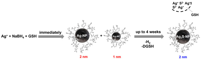
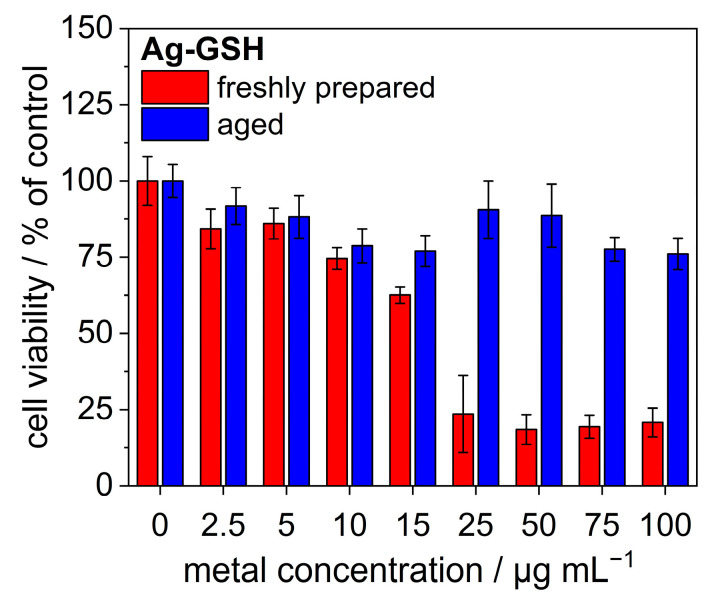
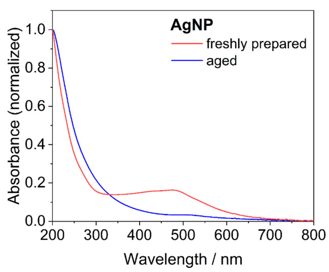
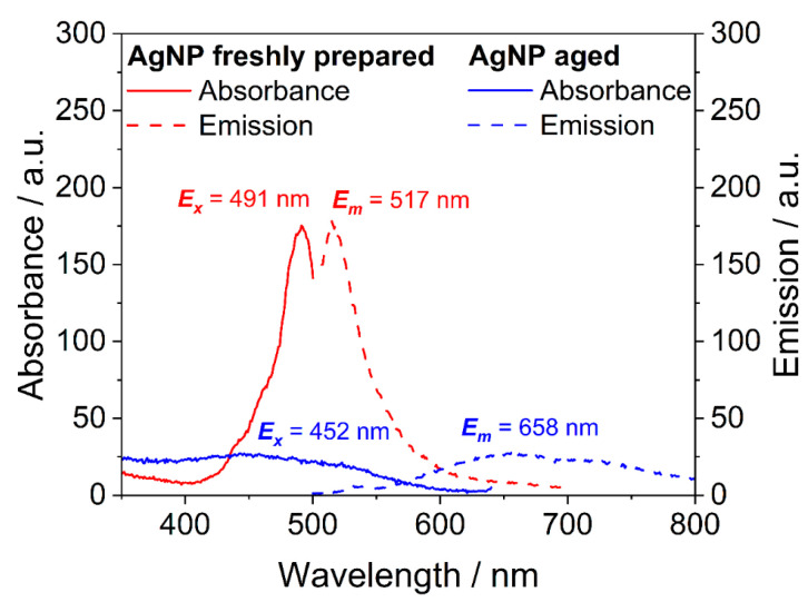
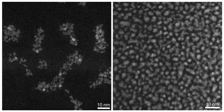
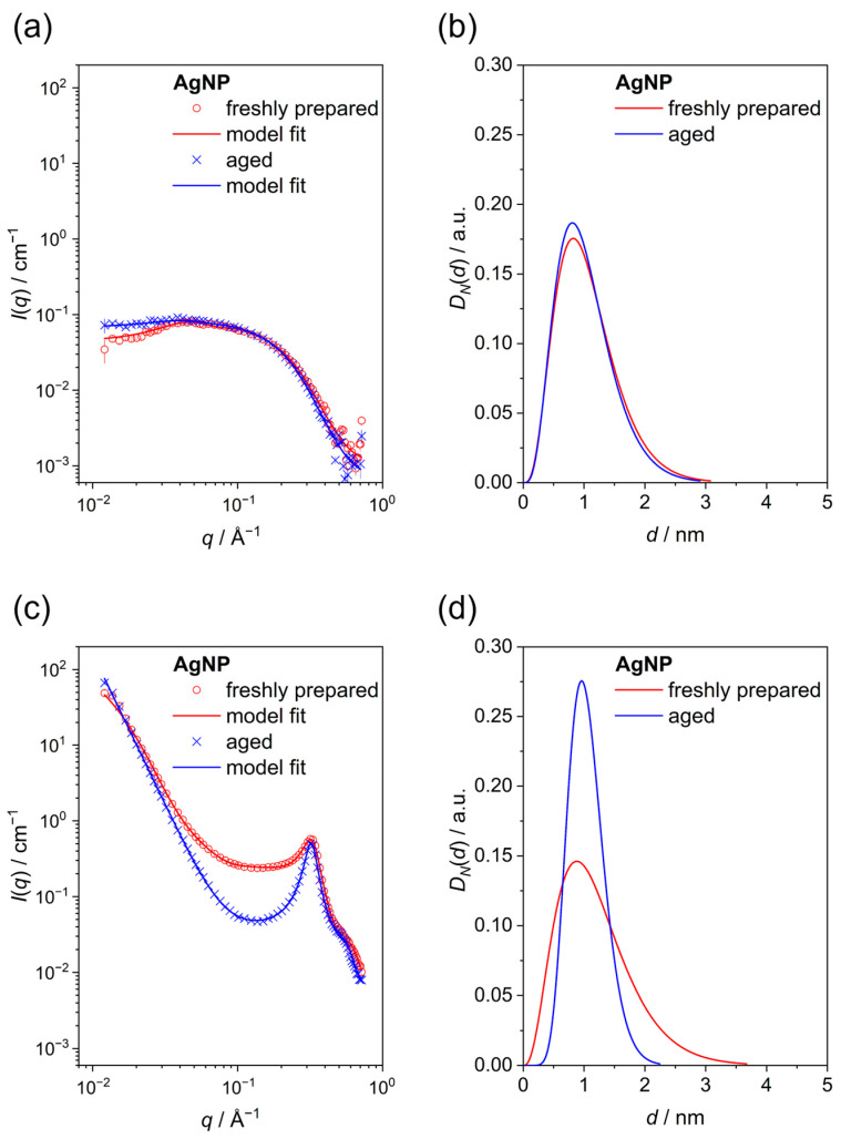
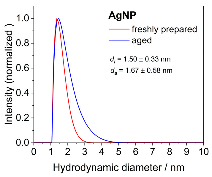
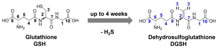
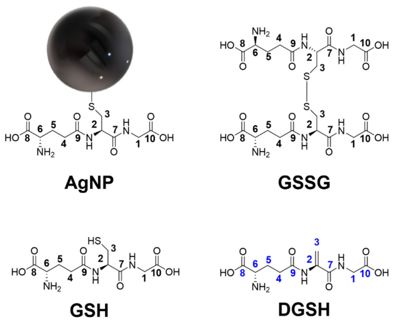
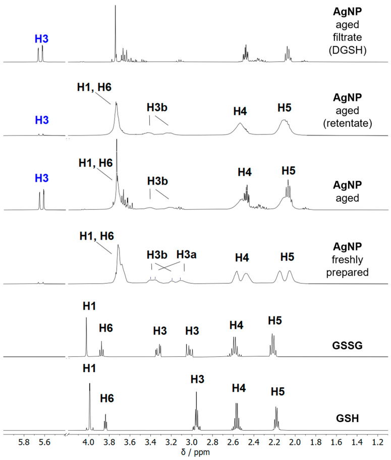
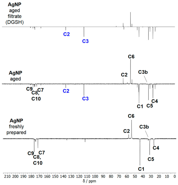
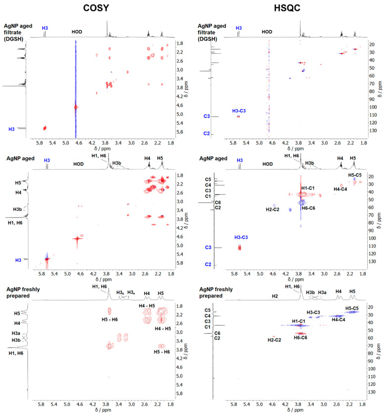
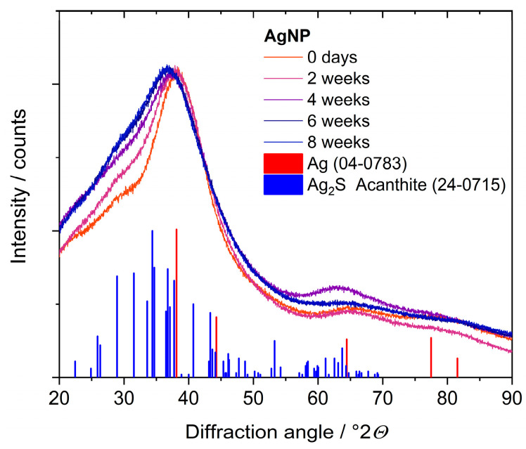
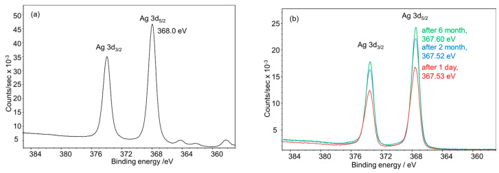
Ultrasmall silver nanoparticles (2 nm) were prepared by reduction with sodium borohydride (NaBH) and stabilized by the ligand glutathione (a tripeptide: glycine-cysteine-glutamic acid). NMR spectroscopy and optical spectroscopy (UV and fluorescence) revealed that these particles initially consist of silver nanoparticles and fluorescing silver nanoclusters, both stabilized by glutathione. Over time, the silver nanoclusters disappear and only the silver nanoparticles remain. Furthermore, the capping ligand glutathione eliminates hydrogen sulfide (HS) from the central cysteine and is released from the nanoparticle surface as tripeptide glycine-dehydroalanine-glutamic acid. Hydrogen sulfide reacts with the silver core to form silver sulfide. After four weeks in dispersion at 4 °C, this process is completed. These processes cannot be detected by transmission electron microscopy (TEM), small-angle X-ray scattering (SAXS), or differential centrifugal sedimentation (DCS) as these methods cannot resolve the mixture of nanoparticles and nanoclusters or the nature of the nanoparticle core. X-ray photoelectron spectroscopy showed the mostly oxidized state of the silver nanoparticle core, Ag(+I), both in freshly prepared and in aged silver nanoparticles. These results demonstrate that ultrasmall nanoparticles can undergo unnoticed changes that considerably affect their chemical, physical, and biological properties. In particular, freshly prepared ultrasmall silver nanoparticles are much more toxic against cells and bacteria than aged particles because of the presence of the silver clusters.
通过硼氢化钠(NaBH)还原制备了超小银纳米颗粒(2纳米),并用配体谷胱甘肽(一种三肽:甘氨酸-半胱氨酸-谷氨酸)进行稳定化处理。核磁共振光谱和光学光谱(紫外和荧光)显示,这些颗粒最初由银纳米颗粒和发荧光的银纳米团簇组成,二者均由谷胱甘肽稳定化。随着时间推移,银纳米团簇消失,仅留下银纳米颗粒。此外,封端配体谷胱甘肽从中心半胱氨酸中消除硫化氢(HS),并以三肽甘氨酸-脱氢丙氨酸-谷氨酸的形式从纳米颗粒表面释放。硫化氢与银核反应形成硫化银。在4℃下分散四周后,该过程完成。这些过程无法通过透射电子显微镜(TEM)、小角X射线散射(SAXS)或差示离心沉降(DCS)检测到,因为这些方法无法分辨纳米颗粒和纳米团簇的混合物或纳米颗粒核心的性质。X射线光电子能谱显示,无论是新制备的还是老化的银纳米颗粒,其核心银大多处于氧化态Ag(+I)。这些结果表明,超小纳米颗粒可能会发生未被注意到的变化,这些变化会显著影响其化学、物理和生物学性质。特别是,由于银团簇的存在,新制备的超小银纳米颗粒对细胞和细菌的毒性比老化颗粒大得多。