Li Yu-Qian, Wang Chun-Sheng, Zhou Jing-Ru, Wang Jia-Ling, Tailaiti Subi, Lin Jia-Ying, Bayina Batesurong, Cao Li-Wei, Ye Jian-Rong
Department of Anesthesiology, The First Affiliated Hospital of Xinjiang Medical University, Urumqi, Xinjiang, China.
Front Immunol. 2024 Aug 29;15:1388120. doi: 10.3389/fimmu.2024.1388120. eCollection 2024.
In this study, the impact of inhibiting the PI3K/AKT/NF-κB pathway on lung oxidative damage induced by cyst fluid was investigated.
Twenty-four mice were randomly assigned to four groups. Three months after inoculation with hydatid cyst segments, mice in group A were treated with intraperitoneal and intratracheal saline injections; mice in group B were administered a caudal vein injection of a PI3K inhibitor, followed by cyst fluid sensitization; mice in group C received an AKT inhibitor via caudal vein, followed by cyst fluid sensitization; and mice in group D were subjected to cyst fluid sensitization without any inhibitor treatment. Cellular changes in lung tissues across all groups were evaluated, including pathological section analysis. Analysis of pulmonary tissue and serum from these mice included the assessment of PI3K/AKT/NF-κB pathway proteins, inflammatory factors, and related mRNA levels.
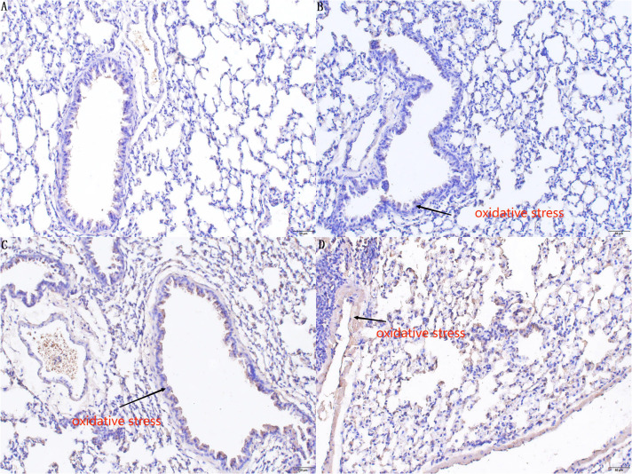
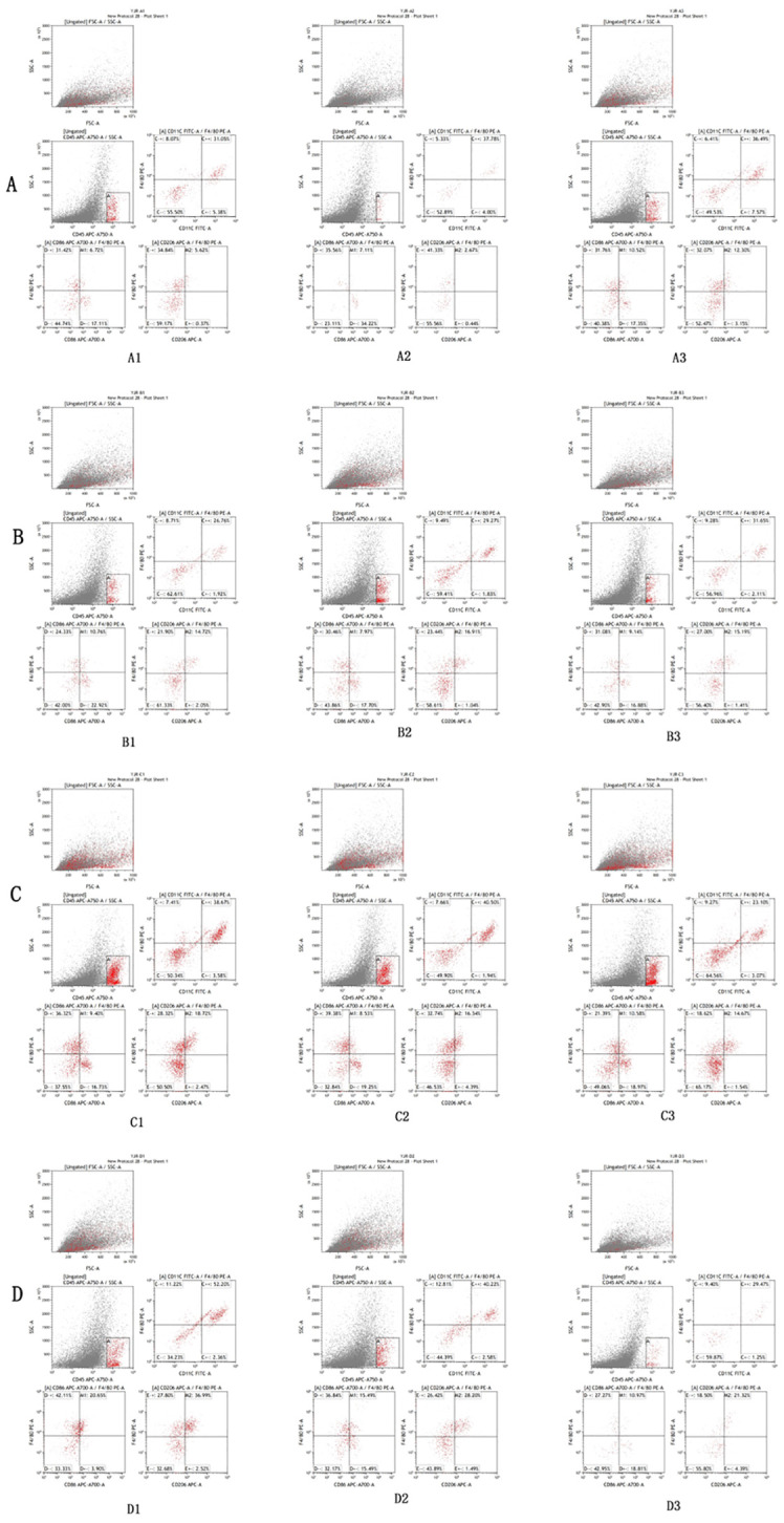
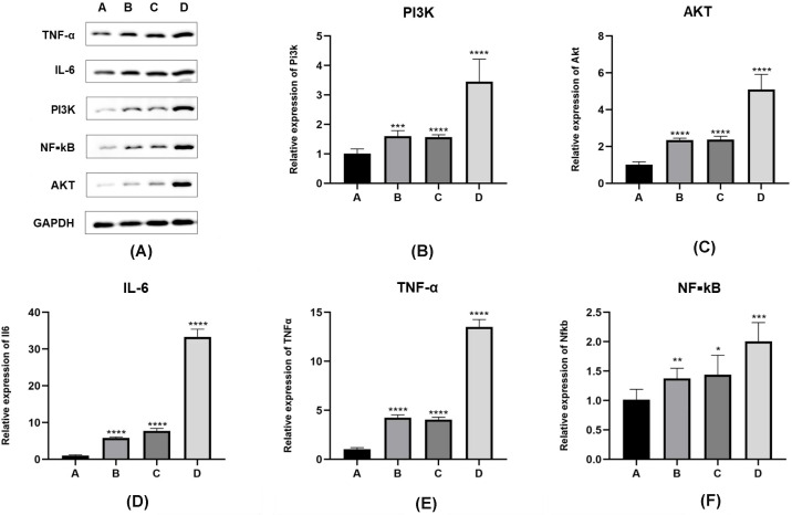
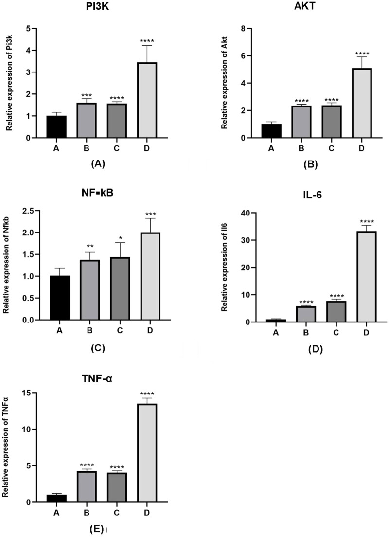
Mice in groups B and C exhibited a higher proportion of M2-type macrophages and significantly lower levels of PI3K/AKT/NF-κB pathway proteins, inflammatory factors (interleukin-6 [IL-6]/tumor necrosis factor-α [TNF-α]), and oxidative markers in lung tissues compared to mice in group D ( 0.05).
Our results in this study indicate that activation of the PI3K/AKT/NF-κB pathway contributed to an increase in the M1 macrophage phenotype, leading to enhanced secretion of peroxidases and inflammatory factors. This mechanism plays a crucial role in the oxidative and inflammatory lung damage associated with allergic reactions to cyst fluid.
本研究旨在探讨抑制PI3K/AKT/NF-κB信号通路对囊液诱导的肺氧化损伤的影响。
将24只小鼠随机分为四组。接种包虫囊肿节段三个月后,A组小鼠经腹腔和气管内注射生理盐水;B组小鼠尾静脉注射PI3K抑制剂,随后进行囊液致敏;C组小鼠尾静脉注射AKT抑制剂,随后进行囊液致敏;D组小鼠进行囊液致敏,不进行任何抑制剂处理。评估所有组肺组织的细胞变化,包括病理切片分析。对这些小鼠的肺组织和血清进行分析,包括评估PI3K/AKT/NF-κB信号通路蛋白、炎症因子及相关mRNA水平。
与D组小鼠相比,B组和C组小鼠肺组织中M2型巨噬细胞比例更高,PI3K/AKT/NF-κB信号通路蛋白、炎症因子(白细胞介素-6 [IL-6]/肿瘤坏死因子-α [TNF-α])和氧化标志物水平显著更低(P<0.05)。
我们在本研究中的结果表明,PI3K/AKT/NF-κB信号通路的激活导致M1巨噬细胞表型增加,从而导致过氧化物酶和炎症因子分泌增加。该机制在与囊液过敏反应相关的肺氧化和炎症损伤中起关键作用。