Palandira Santhoshi P, Falvey Aidan, Carrion Joseph, Zeng Qiong, Chaudhry Saher, Grossman Kira, Turecki Lauren, Nguyen Nha, Brines Michael, Chavan Sangeeta S, Metz Christine N, Al-Abed Yousef, Chang Eric H, Ma Yilong, Eidelberg David, Vo An, Tracey Kevin J, Pavlov Valentin A
The Feinstein Institutes for Medical Research, Northwell Health, Manhasset, NY, USA.
Elmezzi Graduate School of Molecular Medicine, 350 Community Drive, Manhasset, NY 11030, USA.
bioRxiv. 2024 Sep 3:2024.09.02.610840. doi: 10.1101/2024.09.02.610840.
Acute liver injury (ALI) that progresses into acute liver failure (ALF) is a life-threatening condition with an increasing incidence and associated costs. Acetaminophen (N-acetyl-p-aminophenol, APAP) overdosing is among the leading causes of ALI and ALF in the Northern Hemisphere. Brain dysfunction defined as is one of the main diagnostic criteria for ALF. While neuroinflammation and brain metabolic alterations significantly contribute to hepatic encephalopathy, their evaluation at early stages of ALI remained challenging. To provide insights, we utilized post-mortem analysis and non-invasive brain micro positron emission tomography (microPET) imaging of mice with APAP-induced ALI.
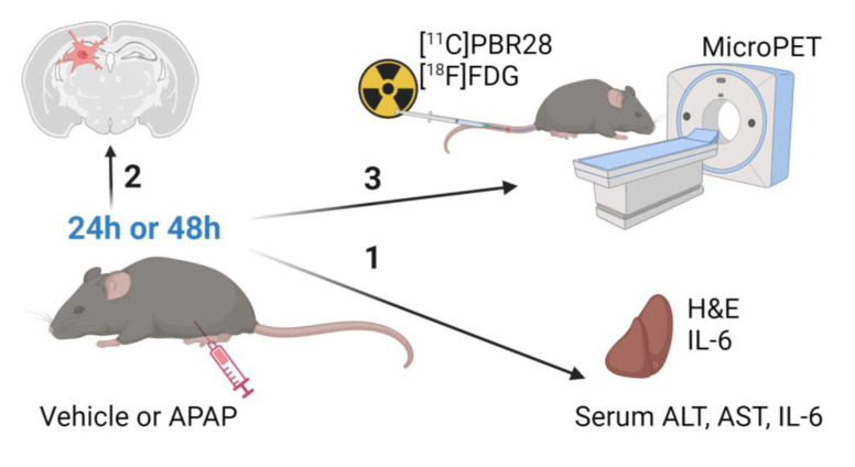
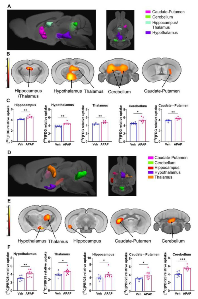
Male C57BL/6 mice were treated with vehicle or APAP (600 mg/kg, i.p.). Serum alanine aminotransferase (ALT), aspartate aminotransferase (AST), liver damage (using H&E staining), hepatic and serum IL-6 levels, and hippocampal IBA1 (using immunolabeling) were evaluated at 24h and 48h. Vehicle and APAP treated animals also underwent microPET imaging utilizing a dual tracer approach, including [C]-peripheral benzodiazepine receptor ([C]PBR28) to assess microglia/astrocyte activation and [F]-fluoro-2-deoxy-2-D-glucose ([F]FDG) to assess energy metabolism. Brain images were pre-processed and evaluated using conjunction and individual tracer uptake analysis.
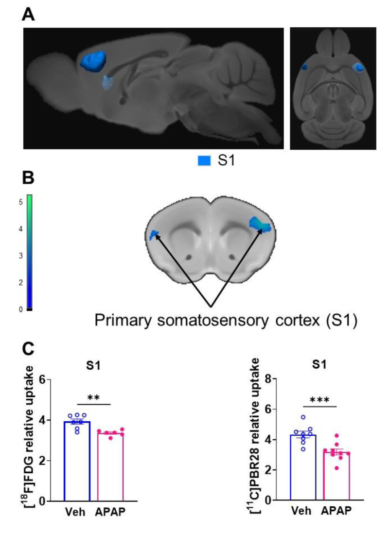
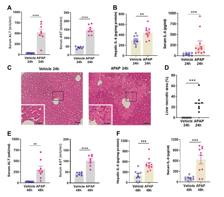
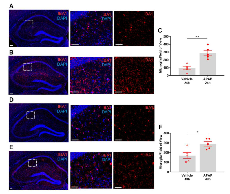
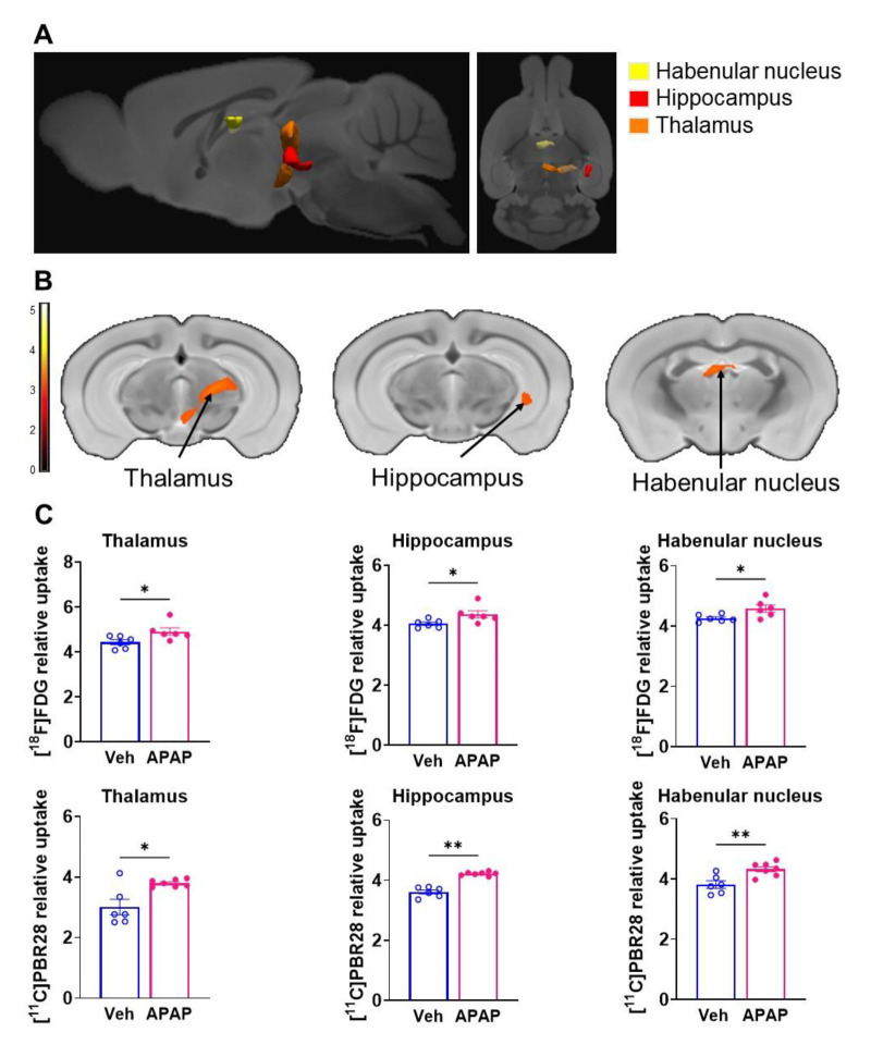
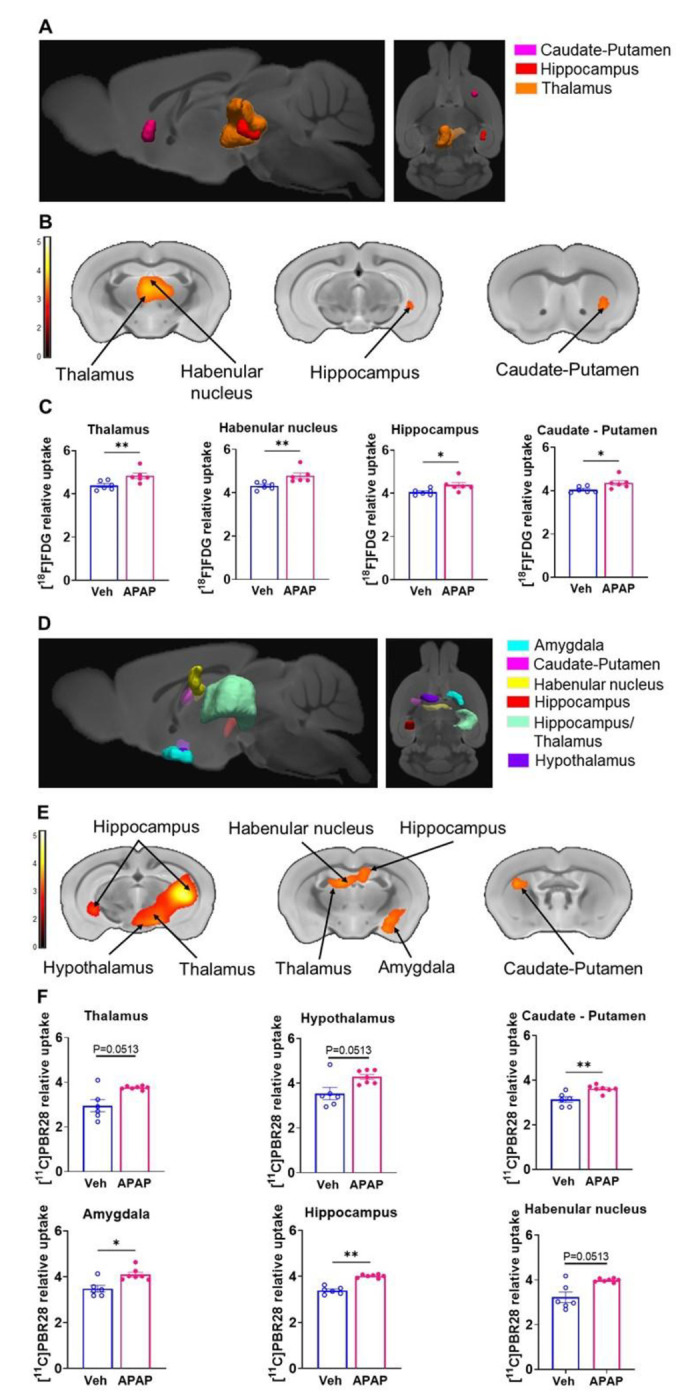
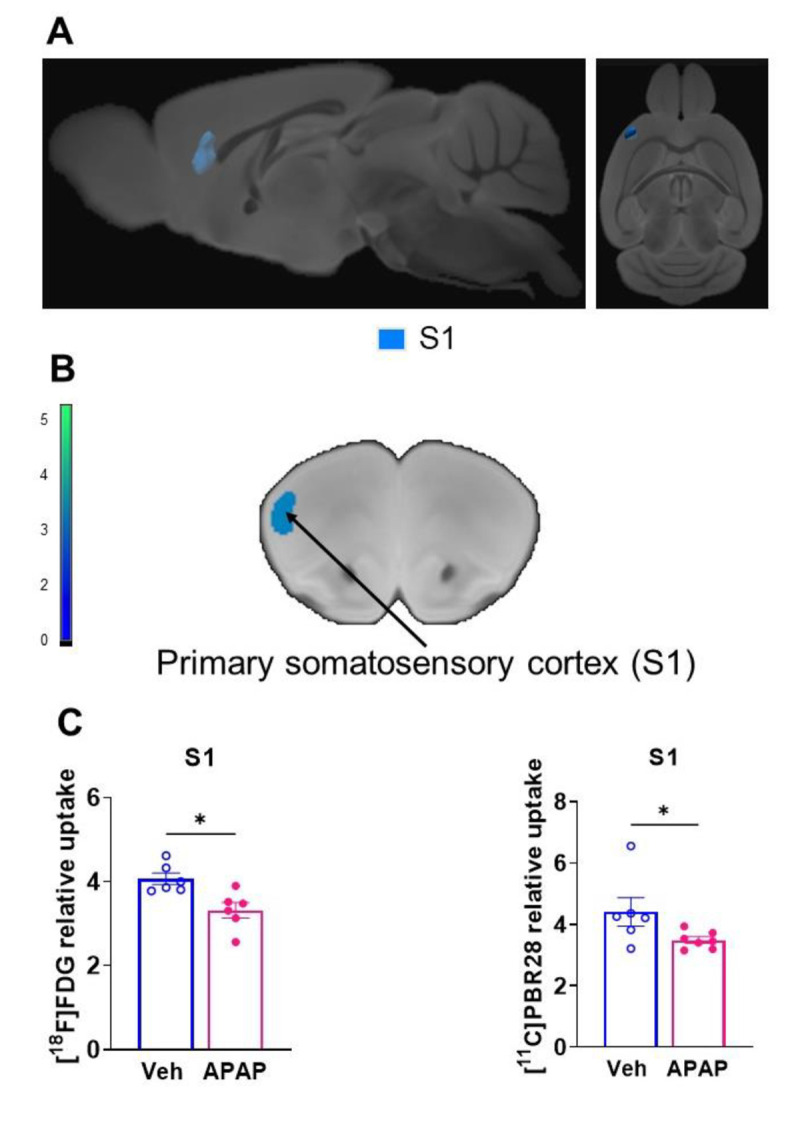
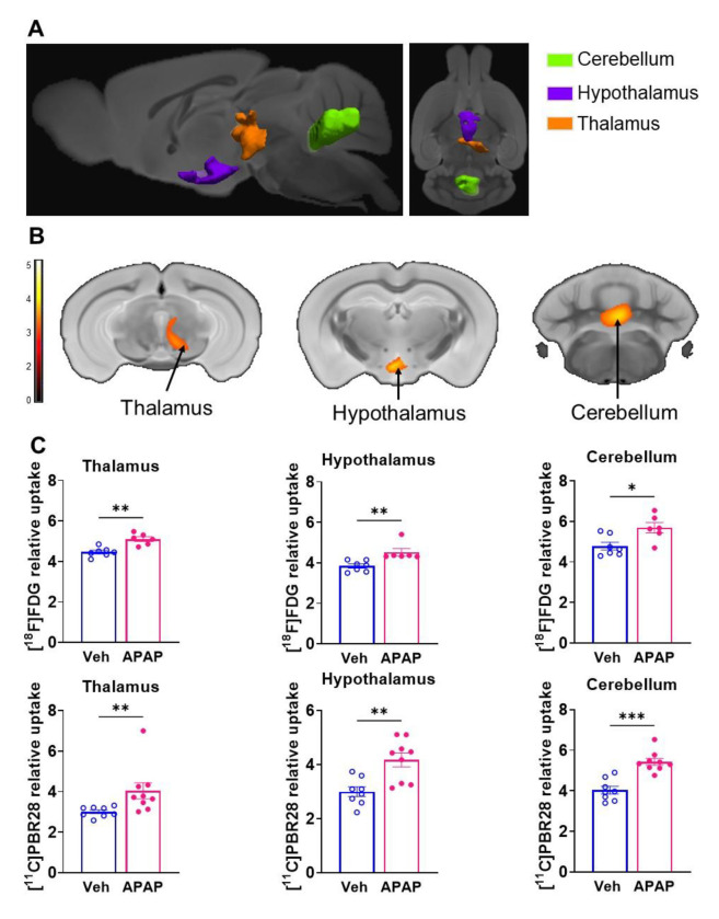
APAP-induced ALI and hepatic and systemic inflammation were detected at 24h and 48h by significantly elevated serum ALT and AST levels, hepatocellular damage, and increased hepatic and serum IL-6 levels. In parallel, increased microglial numbers, indicative for neuroinflammation were observed in the hippocampus of APAP-treated mice. MicroPET imaging revealed overlapping increases in [C]PBR28 and [F]FDG uptake in the hippocampus, thalamus, and habenular nucleus indicating microglial/astroglial activation and increased energy metabolism in APAP-treated mice (vs. vehicle-treated mice) at 24h. Similar significant increases were also found in the hypothalamus, thalamus, and cerebellum at 48h. The individual tracer uptake analyses (APAP vs vehicle) at 24h and 48h confirmed increases in these brain areas and indicated additional tracer- and region-specific effects including hippocampal alterations.
Peripheral manifestations of APAP-induced ALI in mice are associated with brain neuroinflammatory and metabolic alterations at relatively early stages of disease progression, which can be non-invasively evaluated using microPET imaging and conjunction analysis. These findings support further PET-based investigations of brain function in ALI/ALF that may inform timely therapeutic interventions.
进展为急性肝衰竭(ALF)的急性肝损伤(ALI)是一种危及生命的疾病,其发病率和相关成本不断上升。对乙酰氨基酚(N - 乙酰 - 对氨基酚,APAP)过量服用是北半球ALI和ALF的主要原因之一。定义为 的脑功能障碍是ALF的主要诊断标准之一。虽然神经炎症和脑代谢改变显著促成肝性脑病,但其在ALI早期阶段的评估仍然具有挑战性。为了提供见解,我们利用了对APAP诱导的ALI小鼠的尸检分析和非侵入性脑微正电子发射断层扫描(microPET)成像。
雄性C57BL/6小鼠接受载体或APAP(600mg/kg,腹腔注射)治疗。在24小时和48小时评估血清丙氨酸转氨酶(ALT)、天冬氨酸转氨酶(AST)、肝损伤(使用苏木精和伊红染色)、肝脏和血清白细胞介素 - 6水平以及海马IBA1(使用免疫标记)。接受载体和APAP治疗的动物还采用双示踪剂方法进行了microPET成像,包括使用[C] - 外周苯二氮䓬受体([C]PBR28)评估小胶质细胞/星形胶质细胞激活,以及使用[F] - 氟 - 2 - 脱氧 - 2 - D - 葡萄糖([F]FDG)评估能量代谢。脑图像经过预处理,并使用联合分析和单个示踪剂摄取分析进行评估。
在24小时和48小时时,通过血清ALT和AST水平显著升高、肝细胞损伤以及肝脏和血清白细胞介素 - 6水平增加,检测到APAP诱导的ALI以及肝脏和全身炎症。同时,在接受APAP治疗的小鼠海马中观察到小胶质细胞数量增加,这表明存在神经炎症。MicroPET成像显示,在24小时时,接受APAP治疗的小鼠(与接受载体治疗的小鼠相比)海马、丘脑和缰核中[C]PBR28和[F]FDG摄取重叠增加,表明小胶质细胞/星形胶质细胞激活和能量代谢增加。在48小时时,下丘脑、丘脑和小脑中也发现了类似的显著增加。在24小时和48小时进行的单个示踪剂摄取分析(APAP与载体)证实了这些脑区的增加,并表明存在其他示踪剂和区域特异性效应,包括海马改变。
小鼠中APAP诱导的ALI的外周表现与疾病进展相对早期的脑神经炎症和代谢改变相关,这可以使用microPET成像和联合分析进行非侵入性评估。这些发现支持进一步基于PET对ALI/ALF脑功能进行研究,这可能为及时的治疗干预提供依据。