Williams Justin Krish, Ngo Jordan Matthew, Murugupandiyan Abinayaa, Croall Dorothy E, Hartzell H Criss, Schekman Randy
Department of Molecular and Cell Biology, University of California, Berkeley, Berkeley, United States.
Department of Biochemistry, Microbiology and Molecular Biology, University of Maine, Orono, United States.
bioRxiv. 2024 Sep 6:2024.09.05.611512. doi: 10.1101/2024.09.05.611512.
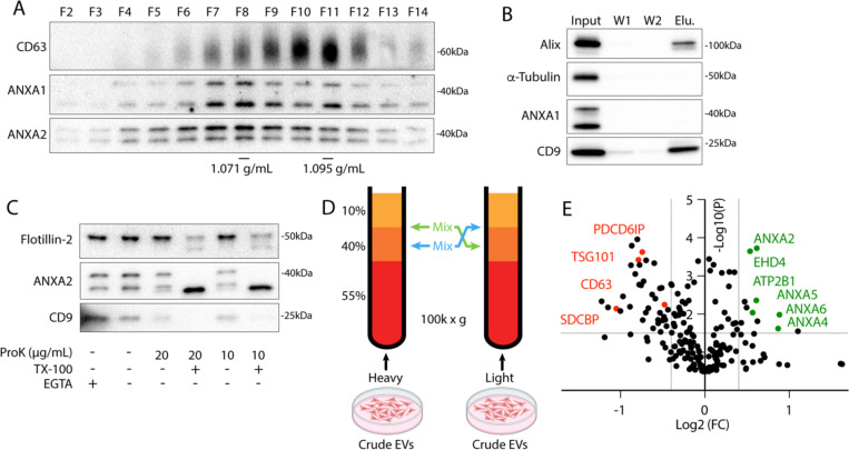
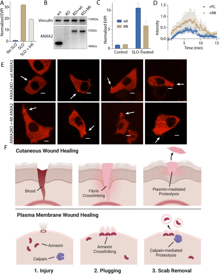
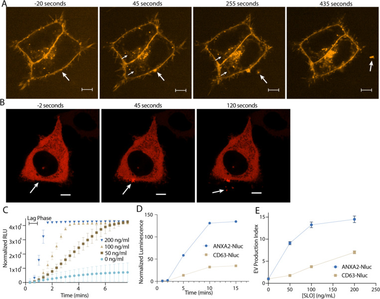
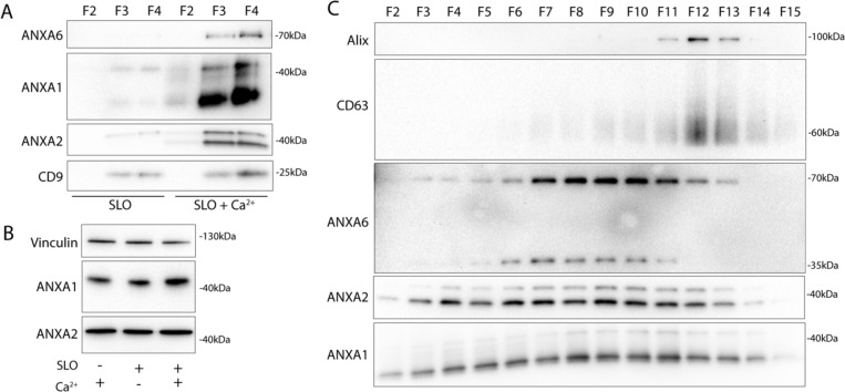
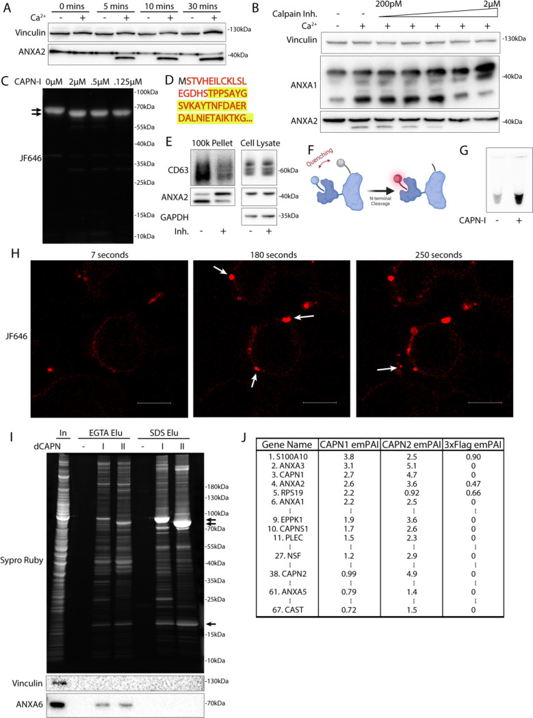
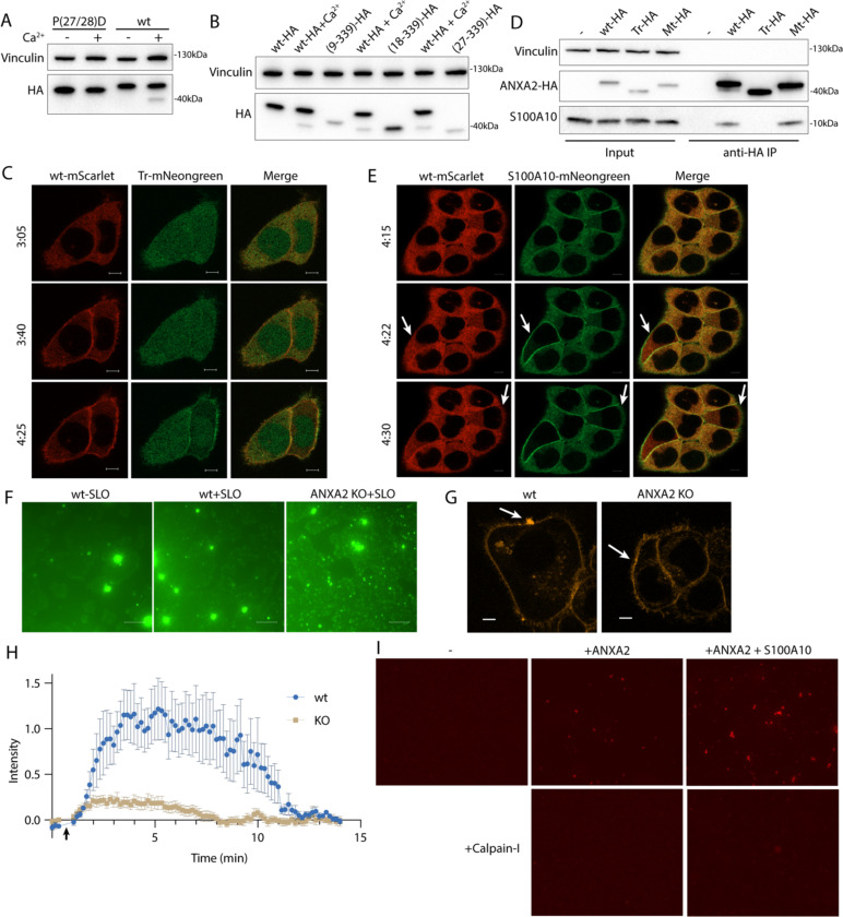
Microvesicles (MVs) are membrane-enclosed, plasma membrane-derived particles released by cells from all branches of life. MVs have utility as disease biomarkers and may participate in intercellular communication; however, physiological processes that induce their secretion are not known. Here, we isolate and characterize annexin-containing MVs and show that these vesicles are secreted in response to the calcium influx caused by membrane damage. The annexins in these vesicles are cleaved by calpains. After plasma membrane injury, cytoplasmic calcium-bound annexins are rapidly recruited to the plasma membrane and form a scab-like structure at the lesion. In a second phase, recruited annexins are cleaved by calpains-1/2, disabling membrane scabbing. Cleavage promotes annexin secretion within MVs. Our data supports a new model of plasma membrane repair, where calpains relax annexin-membrane aggregates in the lesion repair scab, allowing secretion of damaged membrane and annexins as MVs. We anticipate that cells experiencing plasma membrane damage, including muscle and metastatic cancer cells, secrete these MVs at elevated levels.
微囊泡(MVs)是由来自生命各分支的细胞释放的、被膜包裹的、源自质膜的颗粒。微囊泡可用作疾病生物标志物,并可能参与细胞间通讯;然而,诱导其分泌的生理过程尚不清楚。在这里,我们分离并表征了含膜联蛋白的微囊泡,并表明这些囊泡是响应于膜损伤引起的钙内流而分泌的。这些囊泡中的膜联蛋白被钙蛋白酶切割。质膜损伤后,细胞质中与钙结合的膜联蛋白迅速被招募到质膜,并在损伤处形成痂状结构。在第二阶段,被招募的膜联蛋白被钙蛋白酶-1/2切割,使膜结痂失效。切割促进膜联蛋白在微囊泡内的分泌。我们的数据支持了一种新的质膜修复模型,即钙蛋白酶使损伤修复痂中的膜联蛋白-膜聚集体松弛,从而允许受损膜和膜联蛋白作为微囊泡分泌。我们预计,经历质膜损伤的细胞,包括肌肉细胞和转移性癌细胞,会以较高水平分泌这些微囊泡。