Diabetes and Brain Function Unit, Department of Experimental Medical Science, Faculty of Medicine, Lund University, Lund, Sweden.
Wallenberg Centre for Molecular Medicine, Faculty of Medicine, Lund University, Lund, Sweden.
Neurochem Res. 2024 Dec;49(12):3356-3366. doi: 10.1007/s11064-024-04243-4. Epub 2024 Sep 20.
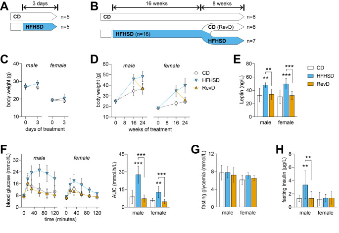
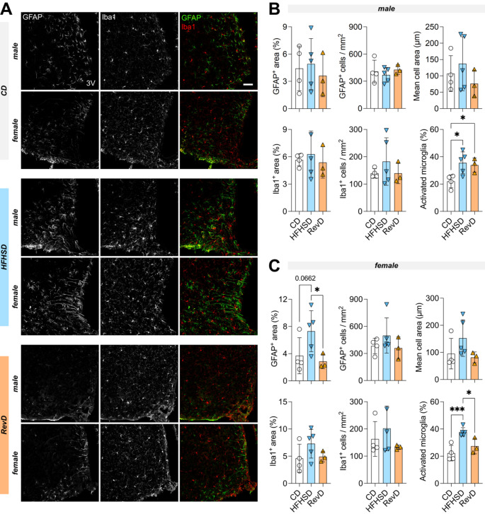
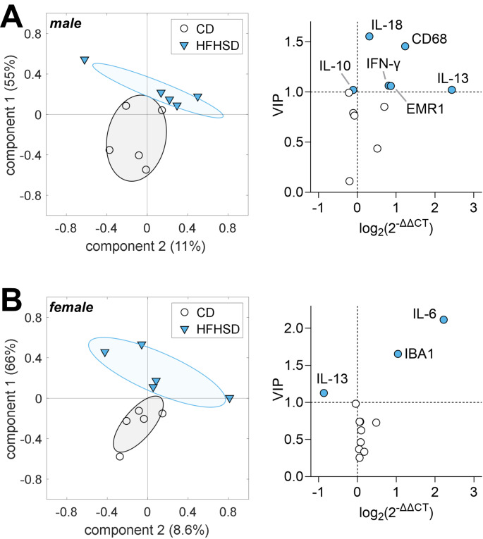
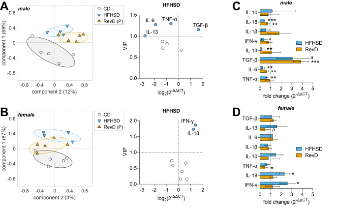
Hypothalamic inflammation underlies diet-induced obesity and diabetes in rodent models. While diet normalization largely allows for recovery from metabolic impairment, it remains unknown whether long-term hypothalamic inflammation induced by obesogenic diets is a reversible process. In this study, we aimed at determining sex specificity of hypothalamic neuroinflammation and gliosis in mice fed a fat- and sugar-rich diet, and their reversibility upon diet normalization. Mice were fed a 60%-fat diet complemented by a 20% sucrose drink (HFHSD) for 3 days or 24 weeks, followed by a third group that had their diet normalized for the last 8 weeks of the study (reverse diet group, RevD). We determined the expression of pro- and anti-inflammatory cytokines, and of the inflammatory cell markers IBA1, CD68, GFAP and EMR1 in the hypothalamus, and analyzed morphology of microglia (IBA-1 cells) and astrocytes (GFAP cells) in the arcuate nucleus. After 3 days of HFHSD feeding, male mice showed over-expression of IL-13, IL-18, IFN-γ, CD68 and EMR1 and reduced expression of IL-10, while females showed increased IL-6 and IBA1 and reduced IL-13, compared to controls. After 24 weeks of HFHSD exposure, male mice showed a general depression in the expression of cytokines, with prominent reduction of TNF-α, IL-6 and IL-13, but increased TGF-β, while female mice showed over-expression of IFN-γ and IL-18. Furthermore, both female and male mice showed some degree of gliosis after HFHSD feeding for 24 weeks. In mice of both sexes, diet normalization after prolonged HFHSD feeding resulted in partial neuroinflammation recovery in the hypothalamus, but gliosis was only recovered in females. In sum, HFHSD-fed mice display sex-specific inflammatory processes in the hypothalamus that are not fully reversible after diet normalization.
下丘脑炎症是啮齿动物模型中饮食诱导肥胖和糖尿病的基础。虽然饮食正常化在很大程度上可以恢复代谢损伤,但肥胖症饮食引起的长期下丘脑炎症是否是一个可逆过程尚不清楚。在这项研究中,我们旨在确定高脂肪和高糖饮食喂养的小鼠下丘脑神经炎症和神经胶质增生的性别特异性,以及饮食正常化后其可逆性。将小鼠用 60%脂肪饮食补充 20%蔗糖饮料(HFHSD)喂养 3 天或 24 周,然后分为第三组,在研究的最后 8 周将其饮食恢复正常(逆转饮食组,RevD)。我们测定了下丘脑促炎和抗炎细胞因子以及炎性细胞标志物 IBA1、CD68、GFAP 和 EMR1 的表达,并分析了弓状核中小胶质细胞(IBA-1 细胞)和星形胶质细胞(GFAP 细胞)的形态。HFHSD 喂养 3 天后,雄性小鼠表现出 IL-13、IL-18、IFN-γ、CD68 和 EMR1 的过度表达以及 IL-10 的表达减少,而雌性小鼠则表现出 IL-6 和 IBA1 的增加以及 IL-13 的减少。HFHSD 暴露 24 周后,雄性小鼠表现出细胞因子表达普遍下降,TNF-α、IL-6 和 IL-13 明显减少,但 TGF-β增加,而雌性小鼠则表现出 IFN-γ和 IL-18 的过度表达。此外,HFHSD 喂养 24 周后,雄性和雌性小鼠均表现出一定程度的神经胶质增生。HFHSD 喂养后,雌雄小鼠的下丘脑神经炎症均有部分恢复,但只有雌性小鼠的神经胶质增生得到恢复。总之,HFHSD 喂养的小鼠表现出下丘脑的性别特异性炎症过程,这些过程在饮食正常化后不能完全恢复。