• 文献检索
  • 文档翻译
  • 深度研究
  • 学术资讯
  • Suppr Zotero 插件Zotero 插件
  • 邀请有礼
  • 套餐&价格
  • 历史记录
应用&插件
Suppr Zotero 插件Zotero 插件浏览器插件Mac 客户端Windows 客户端微信小程序
定价
高级版会员购买积分包购买API积分包
服务
文献检索文档翻译深度研究API 文档MCP 服务
关于我们
关于 Suppr公司介绍联系我们用户协议隐私条款
关注我们

Suppr 超能文献

核心技术专利:CN118964589B侵权必究
粤ICP备2023148730 号-1Suppr @ 2026

文献检索

告别复杂PubMed语法,用中文像聊天一样搜索,搜遍4000万医学文献。AI智能推荐,让科研检索更轻松。

立即免费搜索

文件翻译

保留排版,准确专业,支持PDF/Word/PPT等文件格式,支持 12+语言互译。

免费翻译文档

深度研究

AI帮你快速写综述,25分钟生成高质量综述,智能提取关键信息,辅助科研写作。

立即免费体验

扩展视野 - 一种用于电子显微镜数据交互式可视化的简单方法。

Expanding the field of view - a simple approach for interactive visualisation of electron microscopy data.

机构信息

Department of Biosciences, University of Oslo, Blindernveien 31, PO Box 1041, 0316 Oslo, Norway.

出版信息

J Cell Sci. 2024 Oct 15;137(20). doi: 10.1242/jcs.262198. Epub 2024 Oct 23.

DOI:10.1242/jcs.262198
PMID:39324375
原文链接:https://pmc.ncbi.nlm.nih.gov/articles/PMC11529876/
Abstract

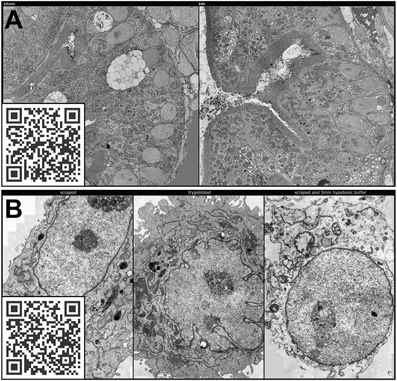
The unparalleled resolving power of electron microscopy is both a blessing and a curse. At 30,000× magnification, 1 µm corresponds to 3 cm in the image and the field of view is only a few micrometres or less, resulting in an inevitable reduction in the spatial data available in an image. Consequently, the gain in resolution is at the cost of loss of the contextual 'reference space', which is crucial for understanding the embedded structures of interest. This problem is particularly pronounced in immunoelectron microscopy, where the detection of a gold particle is crucial for the localisation of specific molecules. The common solution of presenting high-magnification and overview images side by side often insufficiently represents the cellular environment. To address these limitations, we propose here an interactive visualization strategy inspired by digital maps and GPS modules which enables seamless transitions between different magnifications by dynamically linking virtual low magnification overview images with primary high-resolution data. By enabling dynamic browsing, it offers the potential for a deeper understanding of cellular landscapes leading to more comprehensive analysis of the primary ultrastructural data.

摘要

电子显微镜无与伦比的分辨率既是福也是祸。在 30,000×的放大倍数下,1 µm 在图像中对应 3 cm,视场只有几微米或更小,这导致图像中可用的空间数据不可避免地减少。因此,分辨率的提高是以失去上下文“参考空间”为代价的,而参考空间对于理解感兴趣的嵌入结构至关重要。这个问题在免疫电子显微镜中尤为突出,因为金颗粒的检测对于特定分子的定位至关重要。常见的解决方案是并排呈现高倍放大和概览图像,但通常不足以代表细胞环境。为了解决这些限制,我们在这里提出了一种受数字地图和 GPS 模块启发的交互式可视化策略,通过动态链接虚拟低倍概览图像和主要高分辨率数据,实现不同放大倍数之间的无缝过渡。通过实现动态浏览,它有可能更深入地了解细胞景观,从而更全面地分析主要超微结构数据。

https://cdn.ncbi.nlm.nih.gov/pmc/blobs/d389/11529876/4cfc68cd2b7a/joces-137-262198-g7.jpg
https://cdn.ncbi.nlm.nih.gov/pmc/blobs/d389/11529876/418f7c3f8471/joces-137-262198-g1.jpg
https://cdn.ncbi.nlm.nih.gov/pmc/blobs/d389/11529876/6c81c3246bbc/joces-137-262198-g2.jpg
https://cdn.ncbi.nlm.nih.gov/pmc/blobs/d389/11529876/eaa98a37ac5a/joces-137-262198-g3.jpg
https://cdn.ncbi.nlm.nih.gov/pmc/blobs/d389/11529876/b81d1b8271b2/joces-137-262198-g4.jpg
https://cdn.ncbi.nlm.nih.gov/pmc/blobs/d389/11529876/17ce0144ccb3/joces-137-262198-g5.jpg
https://cdn.ncbi.nlm.nih.gov/pmc/blobs/d389/11529876/d1d44135098a/joces-137-262198-g6.jpg
https://cdn.ncbi.nlm.nih.gov/pmc/blobs/d389/11529876/4cfc68cd2b7a/joces-137-262198-g7.jpg

相似文献

1
Expanding the field of view - a simple approach for interactive visualisation of electron microscopy data.扩展视野 - 一种用于电子显微镜数据交互式可视化的简单方法。
J Cell Sci. 2024 Oct 15;137(20). doi: 10.1242/jcs.262198. Epub 2024 Oct 23.
2
Virtual electron microscopy: a simple implementation creating a new paradigm in ultrastructural examination.虚拟电子显微镜:一种简单的实现方式,为超微结构检查创造了新范式。
Int J Surg Pathol. 2011 Oct;19(5):570-5. doi: 10.1177/1066896911421173. Epub 2011 Sep 8.
3
Computer-assisted visualizations of neural networks: expanding the field of view using seamless confocal montaging.神经网络的计算机辅助可视化:使用无缝共聚焦拼接扩展视野
J Neurosci Methods. 2000 Jun 1;98(2):155-63. doi: 10.1016/s0165-0270(00)00200-4.
4
Virtual electron microscopy in cell biology.细胞生物学中的虚拟电子显微镜技术。
Microsc Res Tech. 2011 Mar;74(3):251-6. doi: 10.1002/jemt.20898.
5
Resolution and reproducibility of measures of the diameter of small collagen fibrils by transmission electron microscopy--application to the rabbit corneal stroma.通过透射电子显微镜测量小胶原纤维直径的分辨率和重现性——应用于兔角膜基质
Micron. 2005;36(4):331-43. doi: 10.1016/j.micron.2005.01.003. Epub 2005 Feb 17.
6
Blik is an extensible 3D visualisation tool for the annotation and analysis of cryo-electron tomography data.Blik 是一个可扩展的 3D 可视化工具,用于对冷冻电子断层扫描数据进行注释和分析。
PLoS Biol. 2024 Apr 30;22(4):e3002447. doi: 10.1371/journal.pbio.3002447. eCollection 2024 Apr.
7
Applications and challenges of digital pathology and whole slide imaging.数字病理学与全切片成像的应用及挑战
Biotech Histochem. 2015 Jul;90(5):341-7. doi: 10.3109/10520295.2015.1044566. Epub 2015 May 15.
8
Specular Microscopy镜面显微镜检查
9
An overview of state-of-the-art image restoration in electron microscopy.电子显微镜中最先进的图像恢复技术概述。
J Microsc. 2018 Sep;271(3):239-254. doi: 10.1111/jmi.12716. Epub 2018 Jun 8.
10
Flexible workflows for on-the-fly electron-microscopy single-particle image processing using Scipion.使用 Scipion 实现电子显微镜单颗粒图像处理的灵活工作流程。
Acta Crystallogr D Struct Biol. 2019 Oct 1;75(Pt 10):882-894. doi: 10.1107/S2059798319011860.

引用本文的文献

1
..
Diagnostics (Basel). 2025 Jun 15;15(12):1520. doi: 10.3390/diagnostics15121520.

本文引用的文献

1
Identification of a pharyngeal mucosal lymphoid organ in zebrafish and other teleosts: Tonsils in fish?斑马鱼和其他硬骨鱼咽部黏膜淋巴器官的鉴定:鱼类的扁桃体?
Sci Adv. 2023 Nov 3;9(44):eadj0101. doi: 10.1126/sciadv.adj0101. Epub 2023 Nov 1.
2
OME-Zarr: a cloud-optimized bioimaging file format with international community support.OME-Zarr:具有国际社区支持的云优化生物成像文件格式。
Histochem Cell Biol. 2023 Sep;160(3):223-251. doi: 10.1007/s00418-023-02209-1. Epub 2023 Jul 10.
3
mEMbrain: an interactive deep learning MATLAB tool for connectomic segmentation on commodity desktops.
mEMbrain:一个交互式深度学习 MATLAB 工具,用于在商用台式机上进行连接组分割。
Front Neural Circuits. 2023 Jun 15;17:952921. doi: 10.3389/fncir.2023.952921. eCollection 2023.
4
Interoperable slide microscopy viewer and annotation tool for imaging data science and computational pathology.可互操作的幻灯片显微镜查看器和注释工具,用于成像数据科学和计算病理学。
Nat Commun. 2023 Mar 22;14(1):1572. doi: 10.1038/s41467-023-37224-2.
5
MoBIE: a Fiji plugin for sharing and exploration of multi-modal cloud-hosted big image data.MoBIE:一款用于共享和探索多模态云托管大图像数据的斐济插件。
Nat Methods. 2023 Apr;20(4):475-476. doi: 10.1038/s41592-023-01776-4.
6
Subcellular localization and therapeutic efficacy of polymeric micellar nanoparticles encapsulating bedaquiline for tuberculosis treatment in zebrafish.载有贝达喹啉的聚合物胶束纳米粒用于治疗结核病的亚细胞定位和疗效研究:斑马鱼模型
Biomater Sci. 2023 Mar 14;11(6):2103-2114. doi: 10.1039/d2bm01835g.
7
IMAGE-IN: Interactive web-based multidimensional 3D visualizer for multi-modal microscopy images.IMAGE-IN:用于多模式显微镜图像的交互式基于网络的多维 3D 可视化工具。
PLoS One. 2022 Dec 30;17(12):e0279825. doi: 10.1371/journal.pone.0279825. eCollection 2022.
8
A scalable and modular automated pipeline for stitching of large electron microscopy datasets.用于拼接大型电子显微镜数据集的可扩展和模块化自动流水线。
Elife. 2022 Jul 26;11:e76534. doi: 10.7554/eLife.76534.
9
Nanoparticle entry into cells; the cell biology weak link.纳米颗粒进入细胞;细胞生物学的薄弱环节。
Adv Drug Deliv Rev. 2022 Sep;188:114403. doi: 10.1016/j.addr.2022.114403. Epub 2022 Jun 29.
10
The BioImage Archive - Building a Home for Life-Sciences Microscopy Data.生物影像归档 - 为生命科学显微镜数据构建一个家。
J Mol Biol. 2022 Jun 15;434(11):167505. doi: 10.1016/j.jmb.2022.167505. Epub 2022 Feb 18.