• 文献检索
  • 文档翻译
  • 深度研究
  • 学术资讯
  • Suppr Zotero 插件Zotero 插件
  • 邀请有礼
  • 套餐&价格
  • 历史记录
应用&插件
Suppr Zotero 插件Zotero 插件浏览器插件Mac 客户端Windows 客户端微信小程序
定价
高级版会员购买积分包购买API积分包
服务
文献检索文档翻译深度研究API 文档MCP 服务
关于我们
关于 Suppr公司介绍联系我们用户协议隐私条款
关注我们

Suppr 超能文献

核心技术专利:CN118964589B侵权必究
粤ICP备2023148730 号-1Suppr @ 2026

文献检索

告别复杂PubMed语法,用中文像聊天一样搜索,搜遍4000万医学文献。AI智能推荐,让科研检索更轻松。

立即免费搜索

文件翻译

保留排版,准确专业,支持PDF/Word/PPT等文件格式,支持 12+语言互译。

免费翻译文档

深度研究

AI帮你快速写综述,25分钟生成高质量综述,智能提取关键信息,辅助科研写作。

立即免费体验

径向梯度长骨支架双相复合成型工艺及平台构建研究

Research on Dual-Phase Composite Forming Process and Platform Construction of Radial Gradient Long Bone Scaffold.

作者信息

Zhang Haiguang, Wang Rui, Song Yongteng, Wang Yahao, Hu Qingxi

机构信息

Rapid Manufacturing Engineering Center, School of Mechatronic Engineering and Automation, Shanghai University, Shanghai 200444, China.

Shanghai Key Laboratory of Intelligent Manufacturing and Robotics, Shanghai University, Shanghai 200072, China.

出版信息

Bioengineering (Basel). 2024 Aug 27;11(9):869. doi: 10.3390/bioengineering11090869.

DOI:10.3390/bioengineering11090869
PMID:39329610
原文链接:https://pmc.ncbi.nlm.nih.gov/articles/PMC11428698/
Abstract

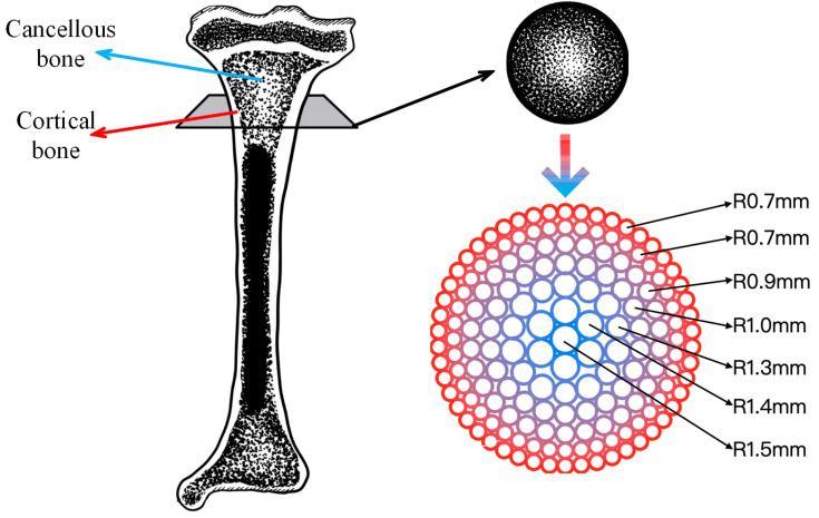
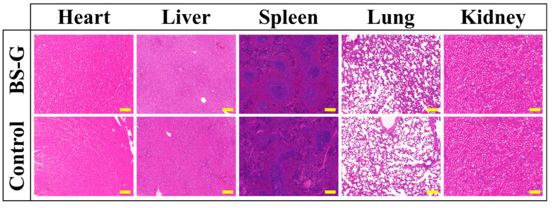
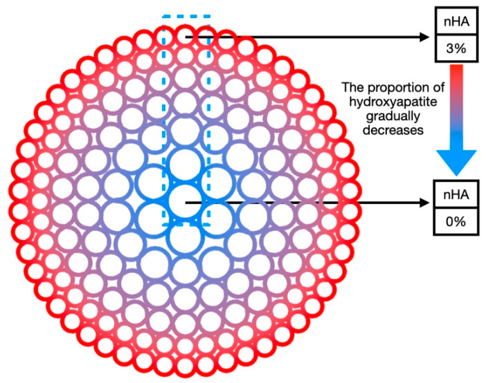
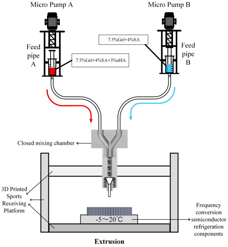
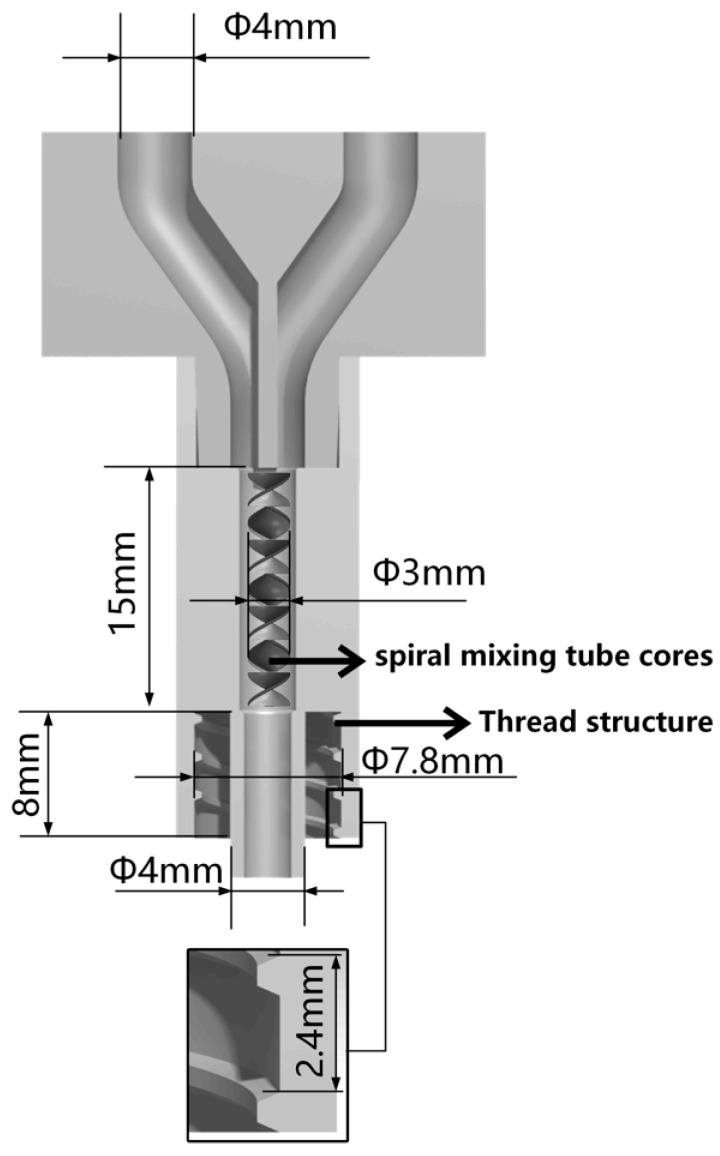
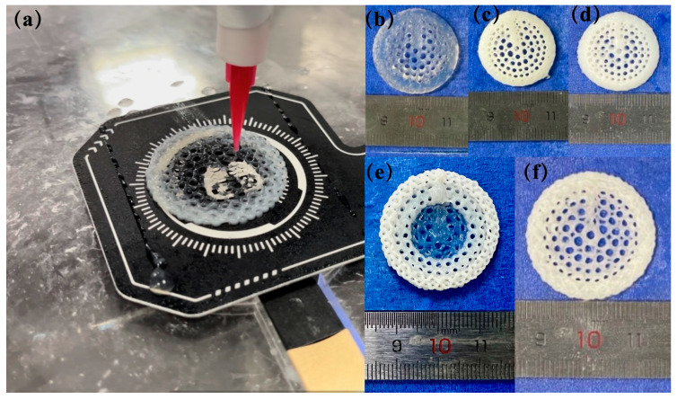
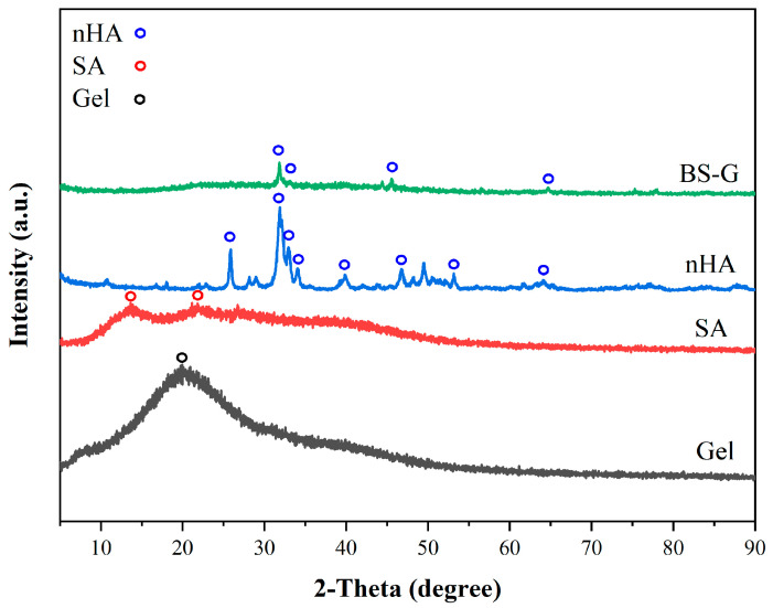
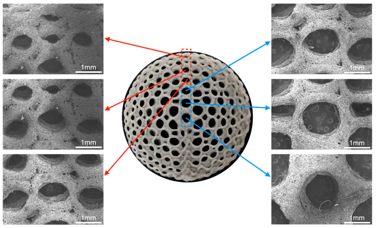
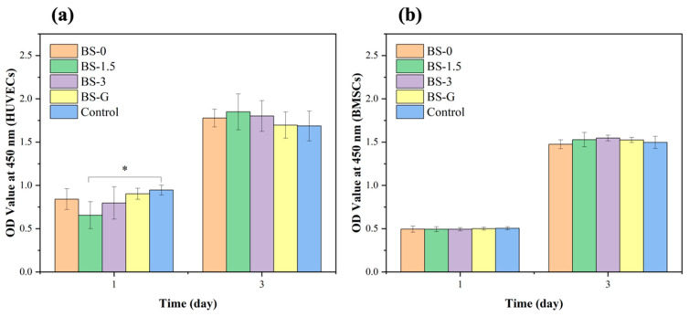
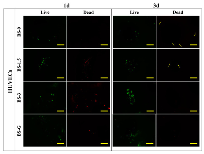
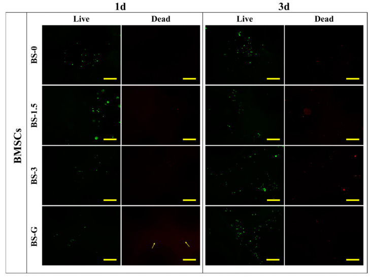
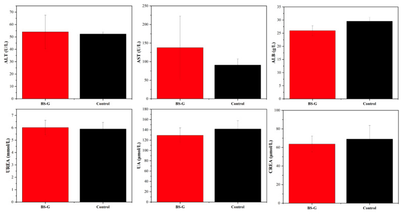
The structure and composition of natural bone show gradient changes. Most bone scaffolds prepared by bone tissue engineering with single materials and structures present difficulties in meeting the needs of bone defect repair. Based on the structure and composition of natural long bones, this study proposed a new bone scaffold preparation technology, the dual-phase composite forming process. Based on the composite use of multiple biomaterials, a bionic natural long bone structure bone scaffold model with bone scaffold pore structure gradient and material concentration gradient changes along the radial direction was designed. Different from the traditional method of using multiple nozzles to achieve material concentration gradient in the scaffold, the dual-phase composite forming process in this study achieved continuous 3D printing preparation of bone scaffolds with gradual material concentration gradient by controlling the speed of extruding materials from two feed barrels into a closed mixing chamber with one nozzle. Through morphological characterization and mechanical property analysis, the results showed that BS-G (radial gradient long bone scaffolds prepared by the dual-phase composite forming process) had obvious pore structure gradient changes and material concentration gradient changes, while BS-T (radial gradient long bone scaffolds prepared by printing three concentrations of material in separate regions) had a discontinuous gradient with obvious boundaries between the parts. The compressive strength of BS-G was 1.00 ± 0.19 MPa, which was higher than the compressive strength of BS-T, and the compressive strength of BS-G also met the needs of bone defect repair. The results of in vitro cell culture tests showed that BS-G had no cytotoxicity. In a Sprague-Dawley rat experimental model, blood tests and key organ sections showed no significant difference between the experimental group and the control group. The prepared BS-G was verified to have good biocompatibility and lays a foundation for the subsequent study of the bone repair effect of radial gradient long bone scaffolds in large animals.

摘要

天然骨的结构和组成呈现出梯度变化。大多数采用单一材料和结构通过骨组织工程制备的骨支架在满足骨缺损修复需求方面存在困难。基于天然长骨的结构和组成,本研究提出了一种新的骨支架制备技术——双相复合成型工艺。基于多种生物材料的复合使用,设计了一种仿生天然长骨结构的骨支架模型,其骨支架孔隙结构梯度和材料浓度梯度沿径向方向变化。与传统使用多个喷嘴在支架中实现材料浓度梯度的方法不同,本研究中的双相复合成型工艺通过控制从两个进料桶向一个带有一个喷嘴的封闭混合腔中挤出材料的速度,实现了具有逐渐材料浓度梯度的骨支架的连续3D打印制备。通过形态表征和力学性能分析,结果表明BS - G(通过双相复合成型工艺制备的径向梯度长骨支架)具有明显的孔隙结构梯度变化和材料浓度梯度变化,而BS - T(通过在不同区域打印三种浓度的材料制备的径向梯度长骨支架)具有不连续的梯度,各部分之间有明显的边界。BS - G的抗压强度为1.00±0.19MPa,高于BS - T的抗压强度,且BS - G的抗压强度也满足骨缺损修复的需求。体外细胞培养测试结果表明BS - G没有细胞毒性。在Sprague - Dawley大鼠实验模型中,血液检测和关键器官切片显示实验组与对照组之间没有显著差异。所制备的BS - G被证实具有良好的生物相容性,为后续研究径向梯度长骨支架在大型动物中的骨修复效果奠定了基础。

https://cdn.ncbi.nlm.nih.gov/pmc/blobs/274b/11428698/b94302c16019/bioengineering-11-00869-g016.jpg
https://cdn.ncbi.nlm.nih.gov/pmc/blobs/274b/11428698/050893de5b56/bioengineering-11-00869-g001.jpg
https://cdn.ncbi.nlm.nih.gov/pmc/blobs/274b/11428698/edf629e8da01/bioengineering-11-00869-g002.jpg
https://cdn.ncbi.nlm.nih.gov/pmc/blobs/274b/11428698/b32841941c49/bioengineering-11-00869-g003.jpg
https://cdn.ncbi.nlm.nih.gov/pmc/blobs/274b/11428698/160d0b2fa54f/bioengineering-11-00869-g004.jpg
https://cdn.ncbi.nlm.nih.gov/pmc/blobs/274b/11428698/c6f80e5aab31/bioengineering-11-00869-g005.jpg
https://cdn.ncbi.nlm.nih.gov/pmc/blobs/274b/11428698/fc4697b0dd2b/bioengineering-11-00869-g006.jpg
https://cdn.ncbi.nlm.nih.gov/pmc/blobs/274b/11428698/b326877fdc6a/bioengineering-11-00869-g007.jpg
https://cdn.ncbi.nlm.nih.gov/pmc/blobs/274b/11428698/58cdd8ed8c84/bioengineering-11-00869-g008.jpg
https://cdn.ncbi.nlm.nih.gov/pmc/blobs/274b/11428698/f69375ddb600/bioengineering-11-00869-g009.jpg
https://cdn.ncbi.nlm.nih.gov/pmc/blobs/274b/11428698/730975bdfc6c/bioengineering-11-00869-g010.jpg
https://cdn.ncbi.nlm.nih.gov/pmc/blobs/274b/11428698/68a40b8a9b97/bioengineering-11-00869-g011.jpg
https://cdn.ncbi.nlm.nih.gov/pmc/blobs/274b/11428698/e3f6d742874b/bioengineering-11-00869-g012.jpg
https://cdn.ncbi.nlm.nih.gov/pmc/blobs/274b/11428698/9c7e33aad5dc/bioengineering-11-00869-g013.jpg
https://cdn.ncbi.nlm.nih.gov/pmc/blobs/274b/11428698/dc2fa98d5bc6/bioengineering-11-00869-g014.jpg
https://cdn.ncbi.nlm.nih.gov/pmc/blobs/274b/11428698/fddecab95010/bioengineering-11-00869-g015.jpg
https://cdn.ncbi.nlm.nih.gov/pmc/blobs/274b/11428698/b94302c16019/bioengineering-11-00869-g016.jpg

相似文献

1
Research on Dual-Phase Composite Forming Process and Platform Construction of Radial Gradient Long Bone Scaffold.径向梯度长骨支架双相复合成型工艺及平台构建研究
Bioengineering (Basel). 2024 Aug 27;11(9):869. doi: 10.3390/bioengineering11090869.
2
Morphological Integrated Preparation Method and Implementation of Inorganic/Organic Dual-Phase Composite Gradient Bionic Bone Scaffold.无机/有机双相复合梯度仿生骨支架的形态学一体化制备方法及实现
3D Print Addit Manuf. 2024 Apr 1;11(2):e607-e618. doi: 10.1089/3dp.2022.0111. Epub 2024 Apr 16.
3
[CYTOCOMPATIBILITY AND PREPARATION OF BONE TISSUE ENGINEERING SCAFFOLD BY COMBINING LOW TEMPERATURE THREE DIMENSIONAL PRINTING AND VACUUM FREEZE-DRYING TECHNIQUES].[低温三维打印与真空冷冻干燥技术相结合制备骨组织工程支架及其细胞相容性]
Zhongguo Xiu Fu Chong Jian Wai Ke Za Zhi. 2016 Mar;30(3):292-7.
4
3D-printed poly(Ɛ-caprolactone) scaffold with gradient mechanical properties according to force distribution in the mandible for mandibular bone tissue engineering.根据下颌骨受力分布具有梯度力学性能的3D打印聚(ε-己内酯)支架用于下颌骨组织工程
J Mech Behav Biomed Mater. 2020 Apr;104:103638. doi: 10.1016/j.jmbbm.2020.103638. Epub 2020 Jan 14.
5
Fabrication and mechanical characterization of 3D printed vertical uniform and gradient scaffolds for bone and osteochondral tissue engineering.用于骨和骨软骨组织工程的 3D 打印垂直均匀和梯度支架的制造和机械特性。
Acta Biomater. 2019 May;90:37-48. doi: 10.1016/j.actbio.2019.03.041. Epub 2019 Mar 21.
6
[Preparation and osteogenesis of acellular dermal matrix/dicalcium phosphate composite scaffold for bone repair].用于骨修复的脱细胞真皮基质/磷酸二钙复合支架的制备及成骨作用
Zhongguo Xiu Fu Chong Jian Wai Ke Za Zhi. 2024 Jun 15;38(6):755-762. doi: 10.7507/1002-1892.202403059.
7
[Preparation and evaluation of tissue engineered osteochondral integration of multi-layered scaffold].多层支架组织工程化骨软骨整合的制备与评价
Zhongguo Xiu Fu Chong Jian Wai Ke Za Zhi. 2018 Apr 15;32(4):434-440. doi: 10.7507/1002-1892.201712038.
8
3D printed polycaprolactone/β-tricalcium phosphate/carbon nanotube composite - Physical properties and biocompatibility.3D打印聚己内酯/β-磷酸三钙/碳纳米管复合材料——物理性能与生物相容性
Heliyon. 2024 Feb 20;10(5):e26071. doi: 10.1016/j.heliyon.2024.e26071. eCollection 2024 Mar 15.
9
[Osteogenesis effect of dynamic mechanical loading on MC3T3-E1 cells in three-dimensional printing biomimetic composite scaffolds].动态机械加载对三维打印仿生复合支架中MC3T3-E1细胞的成骨作用
Zhongguo Xiu Fu Chong Jian Wai Ke Za Zhi. 2018 Apr 15;32(4):448-456. doi: 10.7507/1002-1892.201711091.
10
3D-printed nanohydroxyapatite/methylacrylylated silk fibroin scaffold for repairing rat skull defects.用于修复大鼠颅骨缺损的3D打印纳米羟基磷灰石/甲基丙烯酰化丝素蛋白支架
J Biol Eng. 2024 Mar 21;18(1):22. doi: 10.1186/s13036-024-00416-5.

本文引用的文献

1
Morphological Integrated Preparation Method and Implementation of Inorganic/Organic Dual-Phase Composite Gradient Bionic Bone Scaffold.无机/有机双相复合梯度仿生骨支架的形态学一体化制备方法及实现
3D Print Addit Manuf. 2024 Apr 1;11(2):e607-e618. doi: 10.1089/3dp.2022.0111. Epub 2024 Apr 16.
2
3D printing nacre powder/sodium alginate scaffold loaded with PRF promotes bone tissue repair and regeneration.3D 打印珍珠层粉/海藻酸钠支架负载富血小板纤维蛋白促进骨组织修复与再生。
Biomater Sci. 2024 Apr 30;12(9):2418-2433. doi: 10.1039/d3bm01936e.
3
Tissue engineered periosteum: Fabrication of a gelatin basedtrilayer composite scaffold with biomimetic properties for enhanced bone healing.
组织工程化骨膜:具有仿生特性的明胶基三层复合支架的构建及其在增强骨愈合中的应用
Int J Biol Macromol. 2024 Apr;263(Pt 2):130371. doi: 10.1016/j.ijbiomac.2024.130371. Epub 2024 Feb 27.
4
Influence of structural parameters of 3D-printed triply periodic minimal surface gyroid porous scaffolds on compression performance, cell response, and bone regeneration.3D 打印的三重周期性极小曲面胞状多孔支架结构参数对压缩性能、细胞反应和骨再生的影响。
J Biomed Mater Res B Appl Biomater. 2024 Jan;112(1):e35337. doi: 10.1002/jbm.b.35337. Epub 2023 Oct 5.
5
Towards a New 3Rs Era in the construction of 3D cell culture models simulating tumor microenvironment.迈向模拟肿瘤微环境的3D细胞培养模型构建中的新3R时代。
Front Oncol. 2023 Apr 3;13:1146477. doi: 10.3389/fonc.2023.1146477. eCollection 2023.
6
Fabrication of 3D Bioprinted Bi-Phasic Scaffold for Bone-Cartilage Interface Regeneration.用于骨-软骨界面再生的3D生物打印双相支架的制造。
Biomimetics (Basel). 2023 Feb 21;8(1):87. doi: 10.3390/biomimetics8010087.
7
Triply Periodic Minimal Surfaces Sheet Scaffolds for Tissue Engineering Applications: An Optimization Approach toward Biomimetic Scaffold Design.用于组织工程应用的三重周期极小曲面片状支架:仿生支架设计的优化方法
ACS Appl Bio Mater. 2018 Aug 20;1(2):259-269. doi: 10.1021/acsabm.8b00052. Epub 2018 Aug 9.
8
Fractal Design Boosts Extrusion-Based 3D Printing of Bone-Mimicking Radial-Gradient Scaffolds.分形设计助力基于挤出的骨模拟径向梯度支架3D打印。
Research (Wash D C). 2021 Nov 23;2021:9892689. doi: 10.34133/2021/9892689. eCollection 2021.
9
Advances in Filament Structure of 3D Bioprinted Biodegradable Bone Repair Scaffolds.3D生物打印可生物降解骨修复支架的细丝结构进展
Int J Bioprint. 2021 Oct 13;7(4):426. doi: 10.18063/ijb.v7i4.426. eCollection 2021.
10
Co-culture of BMSCs and HUVECs with simvastatin-loaded gelatin nanosphere/chitosan coating on Mg alloy for osteogenic differentiation and vasculogenesis.载辛伐他汀明胶纳米球/壳聚糖涂层镁合金上共培养骨髓间充质干细胞和人脐静脉内皮细胞的成骨分化和血管生成。
Int J Biol Macromol. 2021 Dec 15;193(Pt B):2021-2028. doi: 10.1016/j.ijbiomac.2021.11.032. Epub 2021 Nov 10.