Department of Electrical Engineering, California Institute of Technology, Pasadena, CA, 91125, USA.
Department of Pathology and Immunology, Washington University School of Medicine, St. Louis, MO, 63110, USA.
Sci Rep. 2024 Sep 27;14(1):22328. doi: 10.1038/s41598-024-73428-2.
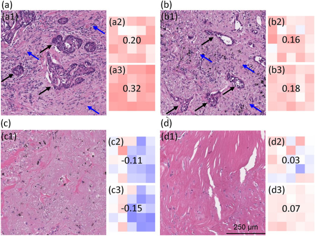
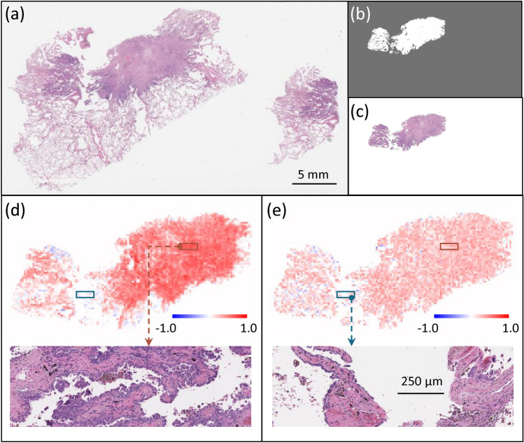
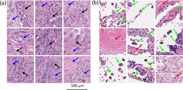
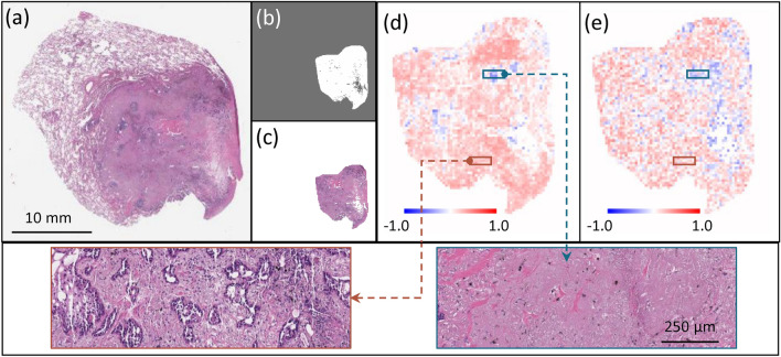
Deep learning-assisted digital pathology has demonstrated the potential to profoundly impact clinical practice, even surpassing human pathologists in performance. However, as deep neural network (DNN) architectures grow in size and complexity, their explainability decreases, posing challenges in interpreting pathology features for broader clinical insights into physiological diseases. To better assess the interpretability of digital microscopic images and guide future microscopic system design, we developed a novel method to study the predictive feature length-scale that underpins a DNN's predictive power. We applied this method to analyze a DNN's capability in predicting brain metastasis from early-stage non-small-cell lung cancer biopsy slides. This study quantifies DNN's attention for brain metastasis prediction, targeting features at both the cellular scale and tissue scale in H&E-stained histological whole slide images. At the cellular scale, the predictive power of DNNs progressively increases with higher resolution and significantly decreases when the resolvable feature length exceeds 5 microns. Additionally, DNN uses more macro-scale features associated with tissue architecture and is optimized when assessing visual fields greater than 41 microns. Our study computes the length-scale requirements for optimal DNN learning on digital whole-slide microscopic images, holding the promise to guide future optical microscope designs in pathology applications and facilitating downstream deep learning analysis.
深度学习辅助数字病理学已经证明了其在临床实践中产生深远影响的潜力,甚至在性能上超越了人类病理学家。然而,随着深度神经网络 (DNN) 架构的规模和复杂性的增加,其可解释性降低,这对解释病理学特征以获得更广泛的生理疾病临床见解提出了挑战。为了更好地评估数字显微镜图像的可解释性,并指导未来显微镜系统的设计,我们开发了一种新的方法来研究支持 DNN 预测能力的预测特征长度尺度。我们将该方法应用于分析 DNN 从早期非小细胞肺癌活检幻灯片中预测脑转移的能力。这项研究量化了 DNN 对脑转移预测的注意力,针对 H&E 染色组织全切片图像中的细胞和组织尺度的特征。在细胞尺度上,DNN 的预测能力随着分辨率的提高而逐步提高,当可分辨特征长度超过 5 微米时,预测能力显著下降。此外,DNN 使用更多与组织架构相关的宏观特征,并在评估大于 41 微米的视场时进行优化。我们的研究计算了数字全切片显微镜图像上 DNN 学习的最佳长度尺度要求,有望指导病理学应用中的未来光学显微镜设计,并促进下游深度学习分析。