Department of Clinical Neuroscience, Division of Eye and Vision, Unit of Optometry, Karolinska Institutet, Stockholm, Sweden.
Department of Molecular Medicine and Surgery, Karolinska Institutet, Sweden.
Invest Ophthalmol Vis Sci. 2024 Oct 1;65(12):4. doi: 10.1167/iovs.65.12.4.
To evaluate the correlation between the macular ganglion cell complex (GCC) thickness measured with manually corrected segmentation and visual function in individuals with chronic Leber hereditary optic neuropathy (LHON).
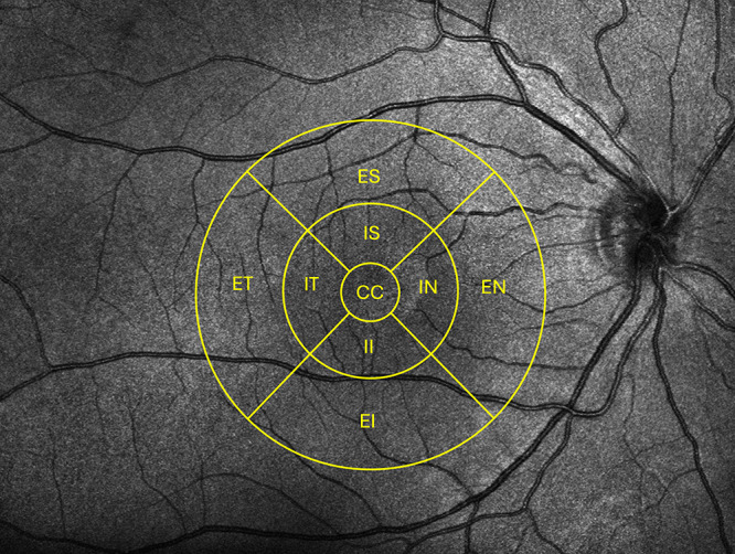
Twenty-six chronic LHON subjects (60% treated with idebenone or Q10) from the Swedish LHON registry were enrolled. Best-corrected visual acuity (BCVA), visual field tests, and optical coherence tomography (OCT) were performed. Visual field was evaluated with the Haag-Streit Octopus 900 with the Esterman test and a custom 30° test. Canon OCT-HS100 scans were exported to the Iowa Reference Algorithm. GCC thickness was obtained after the segmentation was corrected manually in nine macular sectors.
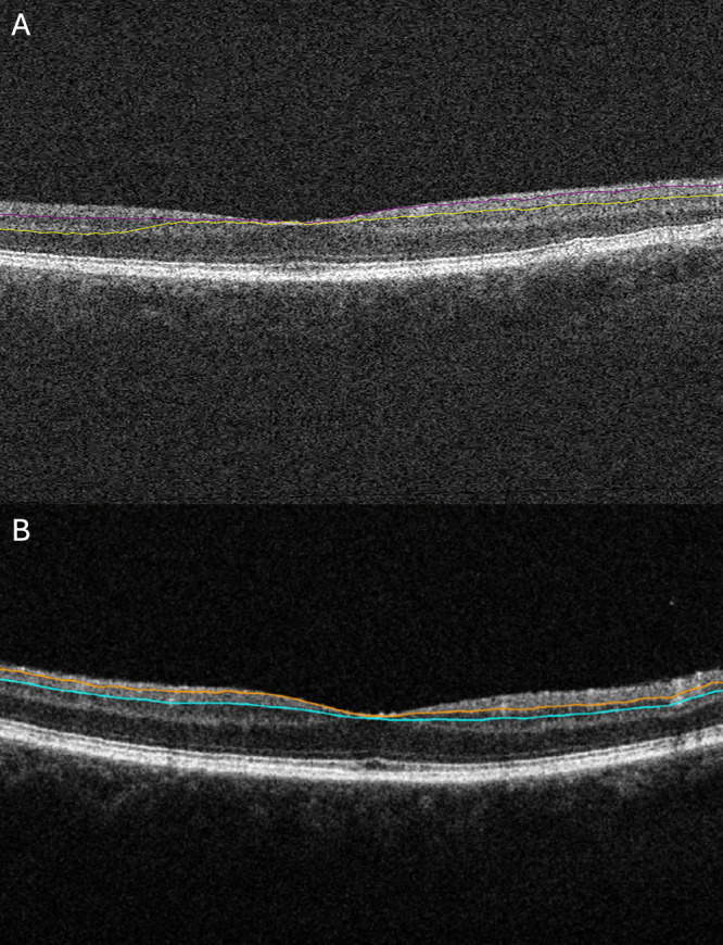
The GCC thickness was overestimated by 16 to 30 µm in different macular sectors with the automated segmentation compared with the corrected (P < 0.001). GCC thickness in all sectors showed significant correlation with all functional parameters. The strongest correlation was seen for the external temporal sector (BCVA: r = 0.604, P < 0.001; mean defect: r = 0.457, P = 0.001; Esterman score: r = 0.421, P = 0.003). No differences were seen between treated and untreated subjects with regard to GCC and visual field scores (P > 0.05), but BCVA was better among treated subjects (P = 0.017).
The corrected GCC thickness showed correlation with visual function in chronic LHON subjects. The frequently occurring segmentation errors in OCT measurements related to chronic LHON can potentially be misleading in monitoring of disease progression and in evaluating the treatment effects. Precise measurements of GCC could serve as a sensitive tool to monitor structural changes in LHON. We therefore emphasize the importance of careful evaluation of the accuracy of OCT segmentation.
评估使用手动校正分割法测量的黄斑神经节细胞复合体(GCC)厚度与慢性莱伯遗传性视神经病变(LHON)患者的视觉功能之间的相关性。
从瑞典 LHON 注册处招募了 26 名患有慢性 LHON 的患者(60%接受 idebenone 或 Q10 治疗)。进行了最佳矫正视力(BCVA)、视野测试和光学相干断层扫描(OCT)。使用 Haag-Streit Octopus 900 进行视野评估,采用 Esterman 测试和定制的 30°测试。将佳能 OCT-HS100 扫描导出到爱荷华参考算法。在手动校正分割后,在九个黄斑区获得了 GCC 厚度。
与校正后相比,自动分割在不同的黄斑区高估了 16 到 30 微米的 GCC 厚度(P<0.001)。所有区的 GCC 厚度与所有功能参数均呈显著相关性。最强的相关性见于外颞区(BCVA:r=0.604,P<0.001;平均缺损:r=0.457,P=0.001;Ester-man 评分:r=0.421,P=0.003)。在 GCC 和视野评分方面,治疗和未治疗的患者之间没有差异(P>0.05),但治疗组的 BCVA 更好(P=0.017)。
校正后的 GCC 厚度与慢性 LHON 患者的视觉功能相关。慢性 LHON 相关的 OCT 测量中经常出现的分割错误可能会对疾病进展的监测和治疗效果的评估产生误导。GCC 的精确测量可以作为监测 LHON 结构变化的敏感工具。因此,我们强调仔细评估 OCT 分割准确性的重要性。