Langhammer Till, Hilbert Kevin, Adolph Dirk, Arolt Volker, Bischoff Sophie, Böhnlein Joscha, Cwik Jan C, Dannlowski Udo, Deckert Jürgen, Domschke Katharina, Evens Ricarda, Fydrich Thomas, Gathmann Bettina, Hamm Alfons O, Heinig Ingmar, Herrmann Martin J, Hollandt Maike, Junghoefer Markus, Kircher Tilo, Koelkebeck Katja, Leehr Elisabeth J, Lotze Martin, Margraf Jürgen, Mumm Jennifer L M, Pittig Andre, Plag Jens, Richter Jan, Roesmann Kati, Ridderbusch Isabelle C, Schneider Silvia, Schwarzmeier Hanna, Seeger Fabian, Siminski Niklas, Straube Thomas, Ströhle Andreas, Szeska Christoph, Wittchen Hans-Ulrich, Wroblewski Adrian, Yang Yunbo, Straube Benjamin, Lueken Ulrike
Department of Psychology, Humboldt-Universität zu Berlin, Berlin, Germany.
Department of Psychology, HMU Health and Medical University Erfurt, Erfurt, Germany.
Mol Psychiatry. 2025 Apr;30(4):1548-1557. doi: 10.1038/s41380-024-02768-2. Epub 2024 Oct 4.
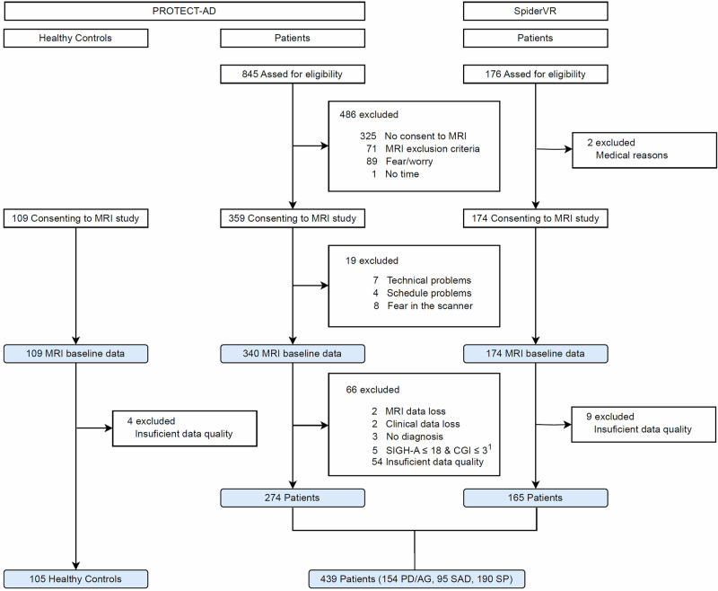
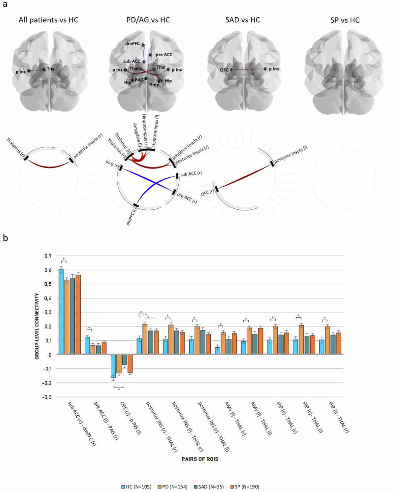
Anxiety disorders (AD) are associated with altered connectivity in large-scale intrinsic brain networks. It remains uncertain how much these signatures overlap across different phenotypes due to a lack of well-powered cross-disorder comparisons. We used resting-state functional magnetic resonance imaging (rsfMRI) to investigate differences in functional connectivity (FC) in a cross-disorder sample of AD patients and healthy controls (HC). Before treatment, 439 patients from two German multicenter clinical trials at eight different sites fulfilling a primary diagnosis of panic disorder and/or agoraphobia (PD/AG, N = 154), social anxiety disorder (SAD, N = 95), or specific phobia (SP, N = 190) and 105 HC underwent an 8 min rsfMRI assessment. We performed categorical and dimensional regions of interest (ROI)-to-ROI analyses focusing on connectivity between regions of the defensive system and prefrontal regulation areas. AD patients showed increased connectivity between the insula and the thalamus compared to controls. This was mainly driven by PD/AG patients who showed increased (insula/hippocampus/amygdala-thalamus) and decreased (dorsomedial prefrontal cortex/periaqueductal gray-anterior cingulate cortex) positive connectivity between subcortical and cortical areas. In contrast, SAD patients showed decreased negative connectivity exclusively in cortical areas (insula-orbitofrontal cortex), whereas no differences were found in SP patients. State anxiety associated with the scanner environment did not explain the FC between these regions. Only PD/AG patients showed pronounced connectivity changes along a widespread subcortical-cortical network, including the midbrain. Dimensional analyses yielded no significant results. The results highlighting categorical differences between ADs at a systems neuroscience level are discussed within the context of personalized neuroscience-informed treatments. PROTECT-AD's registration at NIMH Protocol Registration System: 01EE1402A and German Register of Clinical Studies: DRKS00008743. SpiderVR's registration at ClinicalTrials.gov: NCT03208400.
焦虑症(AD)与大规模内在脑网络的连接性改变有关。由于缺乏有力的跨疾病比较,这些特征在不同表型之间的重叠程度仍不确定。我们使用静息态功能磁共振成像(rsfMRI)来研究AD患者和健康对照(HC)的跨疾病样本中功能连接(FC)的差异。在治疗前,来自德国两个多中心临床试验的439名患者在八个不同地点接受了8分钟的rsfMRI评估,这些患者初步诊断为惊恐障碍和/或广场恐惧症(PD/AG,N = 154)、社交焦虑障碍(SAD,N = 95)或特定恐惧症(SP,N = 190),另有105名HC参与。我们进行了分类和基于感兴趣区域(ROI)到ROI的分析,重点关注防御系统区域与前额叶调节区域之间的连接。与对照组相比,AD患者脑岛与丘脑之间的连接性增加。这主要由PD/AG患者驱动,他们在皮层下和皮层区域之间显示出增加的(脑岛/海马体/杏仁核 - 丘脑)和减少的(背内侧前额叶皮层/导水管周围灰质 - 前扣带回皮层)正性连接。相比之下,SAD患者仅在皮层区域(脑岛 - 眶额皮层)显示出负性连接减少,而SP患者未发现差异。与扫描环境相关的状态焦虑并不能解释这些区域之间的FC。只有PD/AG患者在包括中脑在内的广泛皮层下 - 皮层网络中显示出明显的连接变化。维度分析未得出显著结果。在个性化神经科学指导治疗的背景下,讨论了在系统神经科学水平上突出不同AD之间分类差异的结果。PROTECT - AD在美国国立精神卫生研究所协议注册系统的注册号:01EE1402A,在德国临床研究注册库的注册号:DRKS00008743。SpiderVR在ClinicalTrials.gov的注册号:NCT03208400。