• 文献检索
  • 文档翻译
  • 深度研究
  • 学术资讯
  • Suppr Zotero 插件Zotero 插件
  • 邀请有礼
  • 套餐&价格
  • 历史记录
应用&插件
Suppr Zotero 插件Zotero 插件浏览器插件Mac 客户端Windows 客户端微信小程序
定价
高级版会员购买积分包购买API积分包
服务
文献检索文档翻译深度研究API 文档MCP 服务
关于我们
关于 Suppr公司介绍联系我们用户协议隐私条款
关注我们

Suppr 超能文献

核心技术专利:CN118964589B侵权必究
粤ICP备2023148730 号-1Suppr @ 2026

文献检索

告别复杂PubMed语法,用中文像聊天一样搜索,搜遍4000万医学文献。AI智能推荐,让科研检索更轻松。

立即免费搜索

文件翻译

保留排版,准确专业,支持PDF/Word/PPT等文件格式,支持 12+语言互译。

免费翻译文档

深度研究

AI帮你快速写综述,25分钟生成高质量综述,智能提取关键信息,辅助科研写作。

立即免费体验

基于 MRI T2WI 的放射组学联合 KRAS 基因突变构建模型预测直肠癌肝转移

MRI T2WI-based radiomics combined with KRAS gene mutation constructed models for predicting liver metastasis in rectal cancer.

机构信息

Department of Magnetic Resonance Imaging Diagnostic, The 2nd Affiliated Hospital of Harbin Medical University, Baojian Road, Nangang District, Harbin, 150086, China.

Medical Imaging Center, the Xinjiang Production and Construction Corps Tenth Division Beitun Hospital, Beitun, 836099, China.

出版信息

BMC Med Imaging. 2024 Oct 4;24(1):262. doi: 10.1186/s12880-024-01439-6.

DOI:10.1186/s12880-024-01439-6
PMID:39367333
原文链接:https://pmc.ncbi.nlm.nih.gov/articles/PMC11453062/
Abstract

BACKGROUND

The study aimed to identify the optimal model for predicting rectal cancer liver metastasis (RCLM). This involved constructing various prediction models to aid clinicians in early diagnosis and precise decision-making.

METHODS

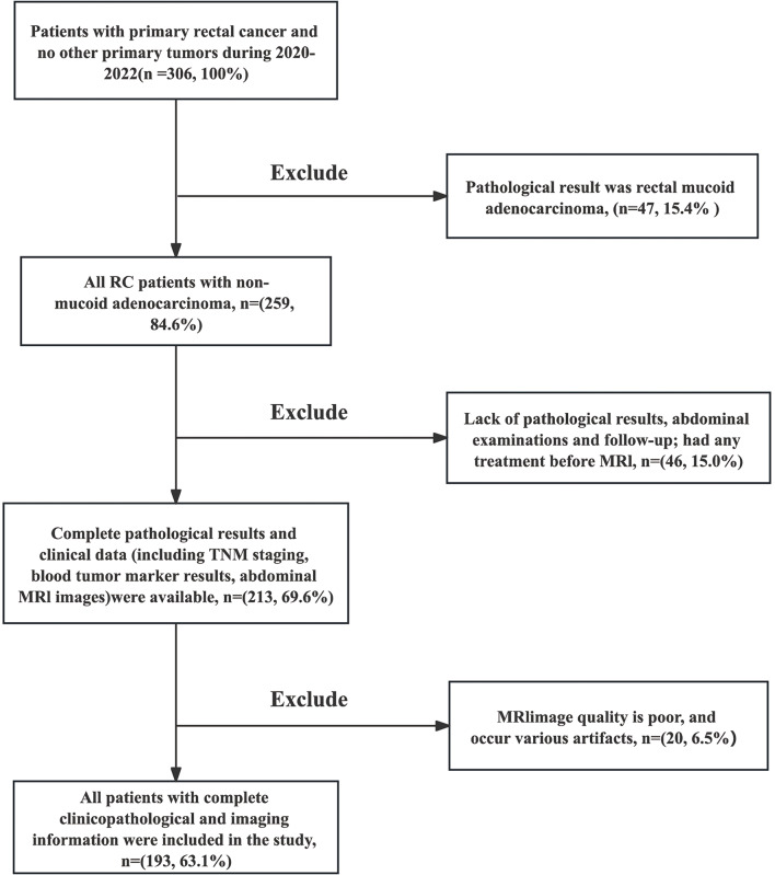
A retrospective analysis was conducted on 193 patients diagnosed with rectal adenocarcinoma were randomly divided into training set (n = 136) and validation set (n = 57) at a ratio of 7:3. The predictive performance of three models was internally validated by 10-fold cross-validation in the training set. Delineation of the tumor region of interest (ROI) was performed, followed by the extraction of radiomics features from the ROI. The least absolute shrinkage and selection operator (LASSO) regression algorithm and multivariate Cox analysis were employed to reduce the dimensionality of radiomics features and identify significant features. Logistic regression was employed to construct three prediction models: clinical, radiomics, and combined models (radiomics + clinical). The predictive performance of each model was assessed and compared.

RESULTS

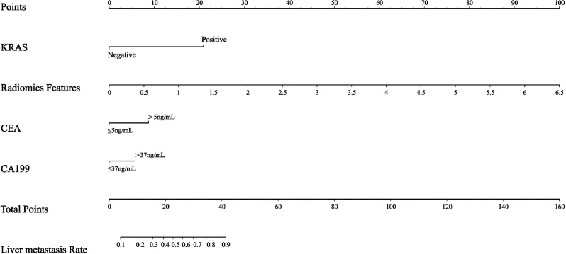
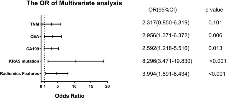
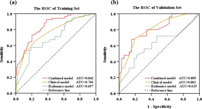
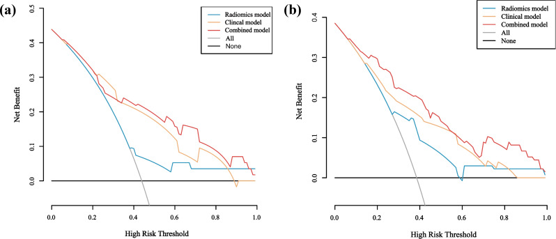
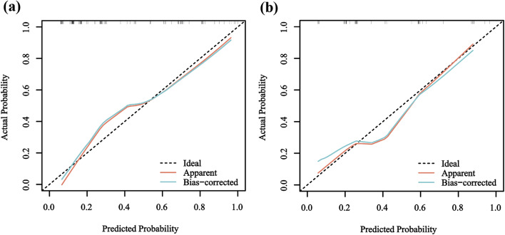
KRAS mutation emerged as an independent predictor of liver metastasis, yielding an odds ratio (OR) of 8.296 (95%CI: 3.471-19.830; p < 0.001). 5 radiomics features will be used to construct radiomics model. The combined model was built by integrating radiomics model with clinical model. In both the training set (AUC:0.842, 95%CI: 0.778-0.907) and the validation set (AUC: 0.805; 95%CI: 0.692-0.918), the AUCs for the combined model surpassed those of the radiomics and clinical models.

CONCLUSIONS

Our study reveals that KRAS mutation stands as an independent predictor of RCLM. The radiomics features based on MR play a crucial role in the evaluation of RCLM. The combined model exhibits superior performance in the prediction of liver metastasis.

CLINICAL TRIAL NUMBER

Not applicable.

摘要

背景

本研究旨在确定预测直肠癌肝转移(RCLM)的最佳模型。这涉及构建各种预测模型,以帮助临床医生进行早期诊断和精确决策。

方法

对 193 例直肠腺癌患者进行回顾性分析,采用 7:3 的比例随机分为训练集(n=136)和验证集(n=57)。在训练集中,通过 10 折交叉验证对内部分类模型的预测性能进行了内部验证。对感兴趣区域(ROI)进行肿瘤勾画,然后从 ROI 中提取放射组学特征。采用最小绝对值收缩和选择算子(LASSO)回归算法和多变量 Cox 分析对放射组学特征进行降维,并识别出显著特征。采用 Logistic 回归构建三个预测模型:临床模型、放射组学模型和联合模型(放射组学+临床模型)。评估并比较了每个模型的预测性能。

结果

KRAS 突变是肝转移的独立预测因素,优势比(OR)为 8.296(95%CI:3.471-19.830;p<0.001)。选取 5 个放射组学特征构建放射组学模型。联合模型是通过整合放射组学模型和临床模型构建的。在训练集(AUC:0.842,95%CI:0.778-0.907)和验证集(AUC:0.805;95%CI:0.692-0.918)中,联合模型的 AUC 均优于放射组学模型和临床模型。

结论

本研究表明,KRAS 突变是 RCLM 的独立预测因素。基于磁共振的放射组学特征在评估 RCLM 中起着重要作用。联合模型在预测肝转移方面具有更好的性能。

临床试验注册号

无。

https://cdn.ncbi.nlm.nih.gov/pmc/blobs/4a93/11453062/49ad6098495f/12880_2024_1439_Fig8_HTML.jpg
https://cdn.ncbi.nlm.nih.gov/pmc/blobs/4a93/11453062/bfaeb9877434/12880_2024_1439_Fig1_HTML.jpg
https://cdn.ncbi.nlm.nih.gov/pmc/blobs/4a93/11453062/a23e6ca65a2b/12880_2024_1439_Fig2_HTML.jpg
https://cdn.ncbi.nlm.nih.gov/pmc/blobs/4a93/11453062/5f07912775b2/12880_2024_1439_Fig3_HTML.jpg
https://cdn.ncbi.nlm.nih.gov/pmc/blobs/4a93/11453062/4fe4521ddf01/12880_2024_1439_Fig4_HTML.jpg
https://cdn.ncbi.nlm.nih.gov/pmc/blobs/4a93/11453062/f13b6b671c9d/12880_2024_1439_Fig5_HTML.jpg
https://cdn.ncbi.nlm.nih.gov/pmc/blobs/4a93/11453062/b5d57a29cde8/12880_2024_1439_Fig6_HTML.jpg
https://cdn.ncbi.nlm.nih.gov/pmc/blobs/4a93/11453062/4d16a38a990f/12880_2024_1439_Fig7_HTML.jpg
https://cdn.ncbi.nlm.nih.gov/pmc/blobs/4a93/11453062/49ad6098495f/12880_2024_1439_Fig8_HTML.jpg

相似文献

1
MRI T2WI-based radiomics combined with KRAS gene mutation constructed models for predicting liver metastasis in rectal cancer.基于 MRI T2WI 的放射组学联合 KRAS 基因突变构建模型预测直肠癌肝转移
BMC Med Imaging. 2024 Oct 4;24(1):262. doi: 10.1186/s12880-024-01439-6.
2
Development and validation of a MRI-based radiomics signature for prediction of KRAS mutation in rectal cancer.基于 MRI 的放射组学特征的建立和验证用于预测直肠癌 KRAS 突变。
Eur Radiol. 2020 Apr;30(4):1948-1958. doi: 10.1007/s00330-019-06572-3. Epub 2020 Jan 15.
3
MRI-based radiomics feature combined with tumor markers to predict TN staging of rectal cancer.基于 MRI 的放射组学特征联合肿瘤标志物预测直肠癌 TN 分期。
J Robot Surg. 2024 May 29;18(1):229. doi: 10.1007/s11701-024-01978-8.
4
MRI-based Radiomics nomogram to detect primary rectal cancer with synchronous liver metastases.基于 MRI 的放射组学列线图检测原发性直肠癌合并同步肝转移。
Sci Rep. 2019 Mar 4;9(1):3374. doi: 10.1038/s41598-019-39651-y.
5
Feasibility of MRI Radiomics for Predicting KRAS Mutation in Rectal Cancer.MRI 影像组学预测直肠癌 KRAS 突变的可行性。
Curr Med Sci. 2020 Dec;40(6):1156-1160. doi: 10.1007/s11596-020-2298-6. Epub 2021 Jan 11.
6
Machine Learning-based Analysis of Rectal Cancer MRI Radiomics for Prediction of Metachronous Liver Metastasis.基于机器学习的直肠癌 MRI 放射组学分析预测肝转移的研究。
Acad Radiol. 2019 Nov;26(11):1495-1504. doi: 10.1016/j.acra.2018.12.019. Epub 2019 Jan 30.
7
[Application of MRI-based Radiomics Models in the Assessment of Hepatic Metastasis of Rectal Cancer].基于MRI的影像组学模型在直肠癌肝转移评估中的应用
Sichuan Da Xue Xue Bao Yi Xue Ban. 2021 Mar;52(2):311-318. doi: 10.12182/20210360202.
8
Diagnostic value of a radiomics model based on CT and MRI for prediction of lateral lymph node metastasis of rectal cancer.基于CT和MRI的影像组学模型对直肠癌侧方淋巴结转移的预测诊断价值
Updates Surg. 2023 Dec;75(8):2225-2234. doi: 10.1007/s13304-023-01618-0. Epub 2023 Aug 9.
9
Preoperative prediction of perineural invasion of rectal cancer based on a magnetic resonance imaging radiomics model: A dual-center study.基于磁共振成像放射组学模型的直肠癌神经周围侵犯的术前预测:一项双中心研究。
World J Gastroenterol. 2024 Apr 28;30(16):2233-2248. doi: 10.3748/wjg.v30.i16.2233.
10
MRI radiomics analysis for predicting preoperative synchronous distant metastasis in patients with rectal cancer.MRI 放射组学分析预测直肠癌患者术前同步远处转移
Eur Radiol. 2019 Aug;29(8):4418-4426. doi: 10.1007/s00330-018-5802-7. Epub 2018 Nov 9.

引用本文的文献

1
Colorectal cancer liver metastases: A radiologic point of view.结直肠癌肝转移:放射学视角
World J Gastrointest Oncol. 2025 Aug 15;17(8):103473. doi: 10.4251/wjgo.v17.i8.103473.
2
Predicting microvascular invasion in solitary hepatocellular carcinoma: a multi-center study integrating clinical, MRI assessments, and radiomics indicators.预测孤立性肝细胞癌中的微血管侵犯:一项整合临床、MRI评估和影像组学指标的多中心研究
Front Oncol. 2025 Feb 19;15:1511260. doi: 10.3389/fonc.2025.1511260. eCollection 2025.

本文引用的文献

1
Estimating age and gender from electrocardiogram signals: A comprehensive review of the past decade.从心电图信号估计年龄和性别:过去十年的综合回顾。
Artif Intell Med. 2023 Dec;146:102690. doi: 10.1016/j.artmed.2023.102690. Epub 2023 Oct 21.
2
Efficacy of fusion imaging for immediate post-ablation assessment of malignant liver neoplasms: A systematic review.融合影像在恶性肝脏肿瘤消融治疗即刻后评估中的疗效:系统评价。
Cancer Med. 2023 Jul;12(13):14225-14251. doi: 10.1002/cam4.6089. Epub 2023 May 16.
3
MRI-based multiregional radiomics for predicting lymph nodes status and prognosis in patients with resectable rectal cancer.
基于磁共振成像的多区域影像组学预测可切除直肠癌患者的淋巴结状态及预后
Front Oncol. 2023 Jan 4;12:1087882. doi: 10.3389/fonc.2022.1087882. eCollection 2022.
4
Dense-PSP-UNet: A neural network for fast inference liver ultrasound segmentation.密集型PSP-UNet:一种用于快速推理肝脏超声分割的神经网络。
Comput Biol Med. 2023 Feb;153:106478. doi: 10.1016/j.compbiomed.2022.106478. Epub 2022 Dec 31.
5
A lightweight neural network with multiscale feature enhancement for liver CT segmentation.一种具有多尺度特征增强的轻量化神经网络,用于肝脏 CT 分割。
Sci Rep. 2022 Aug 19;12(1):14153. doi: 10.1038/s41598-022-16828-6.
6
Practical utility of liver segmentation methods in clinical surgeries and interventions.肝脏分割方法在临床手术和介入中的实际应用。
BMC Med Imaging. 2022 May 24;22(1):97. doi: 10.1186/s12880-022-00825-2.
7
Integrative radiogenomics analysis for predicting molecular features and survival in clear cell renal cell carcinoma.整合放射基因组学分析预测透明细胞肾细胞癌的分子特征和生存。
Aging (Albany NY). 2021 Mar 26;13(7):9960-9975. doi: 10.18632/aging.202752.
8
Global Cancer Statistics 2020: GLOBOCAN Estimates of Incidence and Mortality Worldwide for 36 Cancers in 185 Countries.《全球癌症统计数据 2020:全球 185 个国家和地区 36 种癌症的发病率和死亡率估计》。
CA Cancer J Clin. 2021 May;71(3):209-249. doi: 10.3322/caac.21660. Epub 2021 Feb 4.
9
Prediction of KRAS, NRAS and BRAF status in colorectal cancer patients with liver metastasis using a deep artificial neural network based on radiomics and semantic features.基于影像组学和语义特征的深度人工神经网络预测结直肠癌肝转移患者的KRAS、NRAS和BRAF状态
Am J Cancer Res. 2020 Dec 1;10(12):4513-4526. eCollection 2020.
10
Introduction to Special Issue of Radiology and Imaging of Cancer.癌症放射学与影像学特刊引言
Cancers (Basel). 2020 Sep 18;12(9):2665. doi: 10.3390/cancers12092665.