Department of Orthopaedic Surgery, The Second Affiliated Hospital of Fujian Medical University, 950 Donghai Street, Fengze District, Quanzhou, Fujian, 362000, China.
Department of Orthopaedic Surgery, The First Affiliated Hospital of Fujian Medical University, Fuzhou, 350000, China.
BMC Musculoskelet Disord. 2024 Oct 4;25(1):785. doi: 10.1186/s12891-024-07903-2.
Opportunistic osteoporosis screening, conducted during routine medical examinations such as chest computed tomography (CT), presents a potential solution for early detection. This study aims to investigate the feasibility of utilizing radiomics technology based on chest CT images to screen for opportunistic osteoporosis.
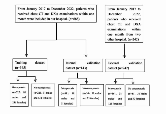
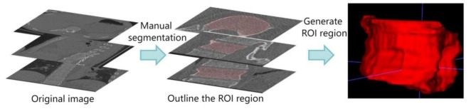
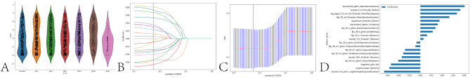
This Study is a Multicenter Retrospective Investigation. Relevant clinical data, including demographics and DXA results, would be collected for each participant. The radiomics analysis in this study focuses on the extraction of features from the 11th or 12th thoracic vertebral bodies from chest CT images. SVM machine learning models would be trained using these radiomic features, with DXA results as the ground truth for osteoporosis classification.
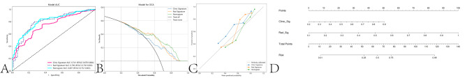
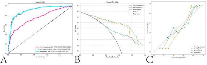
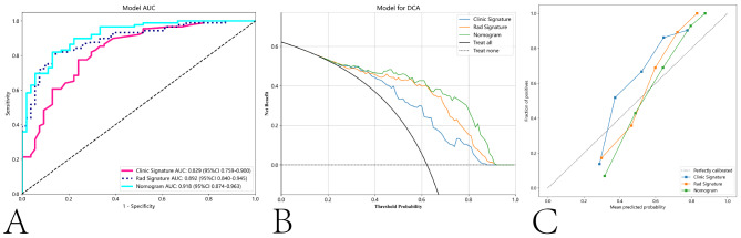
In the training group, Clinical models had an accuracy of 0.684 and an AUC of 0.744, Radiomics models had an accuracy of 0.828 and an AUC of 0.896, Nomogram models had an accuracy of 0.839 and an AUC of 0.901. In the internal validation group, Clinical models had an accuracy of 0.769 and an AUC of 0.829, Radiomics models had an accuracy of 0.832 and an AUC of 0.892, Nomogram models had an accuracy of 0.839 and an AUC of 0.918. In the external validation group, Clinical models had an accuracy of 0.715 and an AUC of 0.741, Radiomics models had an accuracy of 0.777 and an AUC of 0.796, Nomogram models had an accuracy of 0.785 and an AUC of 0.807. In all three datasets, the Nomogram model exhibited a statistically significant difference in screening effectiveness compared to the clinical models.
Our research demonstrates that by leveraging radiomics features extracted from a single thoracic spine using chest CT, and incorporating these features with patient basic information, opportunistic screening for osteoporosis can be achieved.
在常规体检(如胸部 CT)中进行机会性骨质疏松症筛查,为早期发现提供了一种潜在的解决方案。本研究旨在探讨利用胸部 CT 图像的放射组学技术进行机会性骨质疏松症筛查的可行性。
本研究为多中心回顾性研究。将为每位参与者收集相关临床数据,包括人口统计学信息和 DXA 结果。本研究的放射组学分析重点是从胸部 CT 图像中提取第 11 或第 12 胸椎的特征。使用这些放射组学特征训练 SVM 机器学习模型,DXA 结果作为骨质疏松症分类的真实值。
在训练组中,临床模型的准确率为 0.684,AUC 为 0.744,放射组学模型的准确率为 0.828,AUC 为 0.896,列线图模型的准确率为 0.839,AUC 为 0.901。在内部验证组中,临床模型的准确率为 0.769,AUC 为 0.829,放射组学模型的准确率为 0.832,AUC 为 0.892,列线图模型的准确率为 0.839,AUC 为 0.918。在外部验证组中,临床模型的准确率为 0.715,AUC 为 0.741,放射组学模型的准确率为 0.777,AUC 为 0.796,列线图模型的准确率为 0.785,AUC 为 0.807。在所有三个数据集,列线图模型的筛查效果与临床模型相比具有统计学显著差异。
本研究表明,通过利用胸部 CT 提取单个胸椎的放射组学特征,并将这些特征与患者基本信息相结合,可以实现骨质疏松症的机会性筛查。