Morimoto Takayuki, Fujimoto Kenta, Ko Sung-Chul, Nishioka Toshikazu, Tokunaga Hidemori
Department of Neurosurgery, Nara City Hospital, Nara, Japan.
Department of Neurosurgery, Nara Prefecture General Medical Center, Nara, Japan.
Surg Neurol Int. 2024 Sep 27;15:350. doi: 10.25259/SNI_488_2024. eCollection 2024.
Vertebral artery (VA) stump syndrome (VASS) is an embolic source for cerebral infarction (CI) in the posterior circulation after VA occlusion.
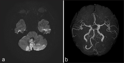
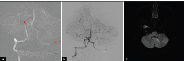
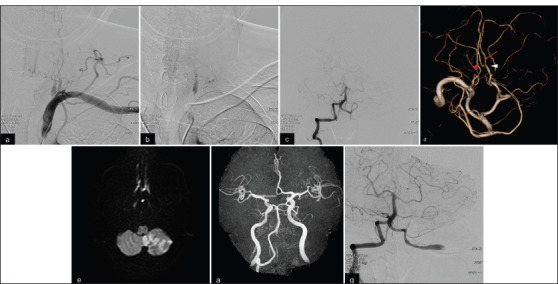
A 63-year-old patient with a history of hypertension presented to our emergent department with dizziness, vomiting, and gait disturbance. Head magnetic resonance imaging (MRI) showed acute CIs in the bilateral cerebellar hemispheres and the vermis. Magnetic resonance angiography revealed patency of the VA and basilar artery. Left subclavian artery digital subtraction angiography (DSA) revealed severe left VA orifice stenosis and collateral flow from the deep cervical artery into the left V2 segment. Right VA angiography showed retrograde flow to the left V4 segment, branching bihemispheric posterior inferior cerebellar artery (PICA), and to-and-flow appearance in the proximal PICA segment and VA. VASS was diagnosed, and conservative treatment with aspirin was administered. Worsened nausea and gait disturbance had developed during hospitalization. MRI revealed an enlarged posterior circulation CI. Follow-up DSA revealed proximal to-and-flow appearance translocation to the proximal V4 segment and poor PICA flow. We performed proximal V4 segment parent artery occlusion (PAO) by endovascular therapy. No recurrence of symptoms or CI was observed. The patient was discharged on day 32 of hospitalization with 1 on the modified Rankin scale.
We reported a rare case of VASS involving bihemispheric PICA. No CI recurrence was observed after performing PAO of the proximal V4 segment. When treating acute cases of bilateral cerebellar CI due to VASS, the contribution of PICA variations should be considered.
椎动脉(VA)残端综合征(VASS)是椎动脉闭塞后后循环脑梗死(CI)的栓子来源。
一名63岁高血压病史患者因头晕、呕吐和步态障碍就诊于我院急诊科。头部磁共振成像(MRI)显示双侧小脑半球和蚓部急性脑梗死。磁共振血管造影显示椎动脉和基底动脉通畅。左锁骨下动脉数字减影血管造影(DSA)显示左椎动脉起始部严重狭窄,有来自颈深动脉至左V2段的侧支血流。右椎动脉血管造影显示血流逆行至左V4段,双半球小脑后下动脉(PICA)分支,以及PICA近端和椎动脉近端的往返血流表现。诊断为VASS,给予阿司匹林保守治疗。住院期间恶心和步态障碍加重。MRI显示后循环脑梗死扩大。随访DSA显示近端往返血流表现移位至近端V4段,PICA血流不佳。我们通过血管内治疗对近端V4段母动脉进行了闭塞(PAO)。未观察到症状或脑梗死复发。患者于住院第32天出院,改良Rankin量表评分为1分。
我们报告了1例罕见的累及双半球PICA的VASS病例。对近端V4段进行PAO后未观察到脑梗死复发。在治疗因VASS导致的双侧小脑急性脑梗死病例时,应考虑PICA变异的影响。