Department of Radiology, Nanfang Hospital, Southern Medical University, Guangzhou, China.
Department of Radiology, The Tenth Affiliated Hospital of Southern Medical University (Dongguan People's Hospital), Dongguan, China.
Technol Cancer Res Treat. 2024 Jan-Dec;23:15330338241289474. doi: 10.1177/15330338241289474.
To assess the diagnostic performance of FFDM-based and DBT-based radiomics models to differentiate breast phyllodes tumors from fibroadenomas.
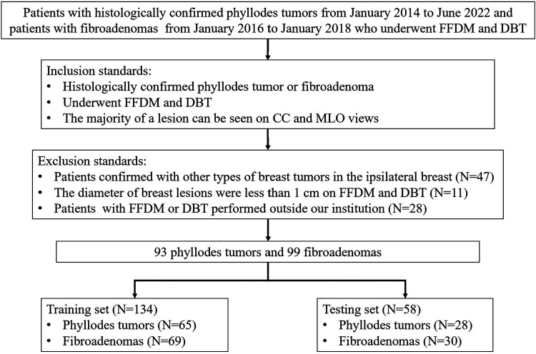
192 patients (93 phyllodes tumors and 99 fibroadenomas) who underwent mammography were retrospectively enrolled. Radiomic features were respectively extracted from FFDM and the clearest slice of DBT images. A least absolute shrinkage and selection operator (LASSO) regression was used to select radiomics features. A combined model was constructed by radiomics and radiological signatures. Machine learning classification was done using logistic regression based on radiomics or radiological signatures (clinical model). Four radiologists were tested on phyllodes tumors and fibroadenomas with and without optimal model assistance. The area under the receiver operating characteristic (ROC) curve (AUC) was computed to assess the performance of each model or radiologist. The Delong test and McNemar's test were performed to compare the performance.
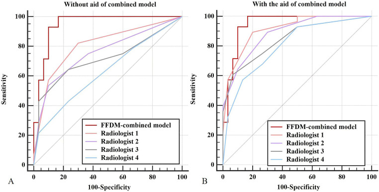
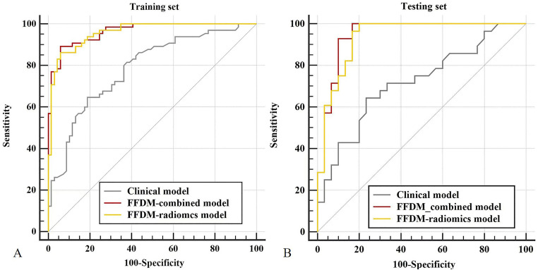
The combined model yielded the highest performance with an AUC of 0.948 (95%CI: 0.889-1.000) in the testing set, slightly higher than the FFDM-radiomics model (AUC of 0.937, 95%CI: 0.841-0.984) and the DBT-radiomics model (AUC of 0.860, 95%CI: 0.742-0.936) and significantly superior to the clinical model (AUC of 0.719, 95%CI: 0.585-0.829). With the combined model aid, the AUCs of four radiologists were improved from 0.808 to 0.914 (=0.079), 0.759 to 0.888 (=0.015), 0.717 to 0.846 (=0.004), and 0.629 to 0.803 (=0.001).
Radiomics analysis based on FFDM and DBT shows promise in differentiating phyllodes tumors from fibroadenomas.
评估基于全数字化乳腺摄影(FFDM)和数字乳腺断层合成(DBT)的放射组学模型对乳腺叶状肿瘤与纤维腺瘤的鉴别诊断性能。
回顾性纳入 192 例(93 例叶状肿瘤和 99 例纤维腺瘤)接受乳腺摄影的患者。分别从 FFDM 和 DBT 最清晰层面图像中提取放射组学特征。采用最小绝对值收缩和选择算子(LASSO)回归筛选放射组学特征。通过放射组学和影像学特征(临床模型)构建联合模型。基于放射组学或影像学特征(临床模型),采用逻辑回归进行机器学习分类。四位放射科医生在有无最佳模型辅助的情况下对叶状肿瘤和纤维腺瘤进行诊断。计算受试者工作特征(ROC)曲线下面积(AUC)以评估每个模型或放射科医生的性能。采用 Delong 检验和 McNemar 检验比较性能。
联合模型在测试集中的 AUC 为 0.948(95%CI:0.889-1.000),性能最高,略高于 FFDM 放射组学模型(AUC 为 0.937,95%CI:0.841-0.984)和 DBT 放射组学模型(AUC 为 0.860,95%CI:0.742-0.936),明显优于临床模型(AUC 为 0.719,95%CI:0.585-0.829)。在联合模型辅助下,四位放射科医生的 AUC 从 0.808 提高到 0.914(=0.079)、0.759 提高到 0.888(=0.015)、0.717 提高到 0.846(=0.004)和 0.629 提高到 0.803(=0.001)。
基于 FFDM 和 DBT 的放射组学分析在鉴别叶状肿瘤与纤维腺瘤方面具有潜力。