• 文献检索
  • 文档翻译
  • 深度研究
  • 学术资讯
  • Suppr Zotero 插件Zotero 插件
  • 邀请有礼
  • 套餐&价格
  • 历史记录
应用&插件
Suppr Zotero 插件Zotero 插件浏览器插件Mac 客户端Windows 客户端微信小程序
定价
高级版会员购买积分包购买API积分包
服务
文献检索文档翻译深度研究API 文档MCP 服务
关于我们
关于 Suppr公司介绍联系我们用户协议隐私条款
关注我们

Suppr 超能文献

核心技术专利:CN118964589B侵权必究
粤ICP备2023148730 号-1Suppr @ 2026

文献检索

告别复杂PubMed语法,用中文像聊天一样搜索,搜遍4000万医学文献。AI智能推荐,让科研检索更轻松。

立即免费搜索

文件翻译

保留排版,准确专业,支持PDF/Word/PPT等文件格式,支持 12+语言互译。

免费翻译文档

深度研究

AI帮你快速写综述,25分钟生成高质量综述,智能提取关键信息,辅助科研写作。

立即免费体验

用于评估强直性脊柱炎患者影像学损伤的最佳深度学习模型。

An optimal deep learning model for the scoring of radiographic damage in patients with ankylosing spondylitis.

作者信息

Chen Yen-Ju, Chen Der-Yuan, Lan Haw-Chang, Huang An-Chih, Chen Yi-Hsing, Huang Wen-Nan, Chen Hsin-Hua

机构信息

Division of Allergy, Immunology and Rheumatology, Department of Internal Medicine, Taichung Veterans General Hospital, Taichung, Taiwan.

Institute of Clinical Medicine, National Yang Ming University, Taipei, Taiwan.

出版信息

Ther Adv Musculoskelet Dis. 2024 Oct 7;16:1759720X241285973. doi: 10.1177/1759720X241285973. eCollection 2024.

DOI:10.1177/1759720X241285973
PMID:39381056
原文链接:https://pmc.ncbi.nlm.nih.gov/articles/PMC11459512/
Abstract

BACKGROUND

Detecting vertebral structural damage in patients with ankylosing spondylitis (AS) is crucial for understanding disease progression and in research settings.

OBJECTIVES

This study aimed to use deep learning to score the modified Stoke Ankylosing Spondylitis Spinal Score (mSASSS) using lateral X-ray images of the cervical and lumbar spine in patients with AS in Asian populations.

DESIGN

A deep learning model was developed to automate the scoring of mSASSS based on X-ray images.

METHODS

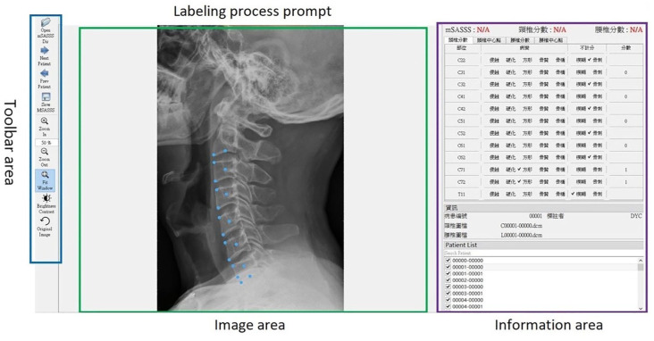
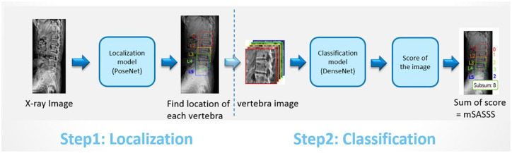
We enrolled patients with AS at a tertiary medical center in Taiwan from August 1, 2001 to December 30, 2020. A localization module was used to locate the vertebral bodies in the images of the cervical and lumbar spine. Images were then extracted from these localized points and fed into a classification module to determine whether common lesions of AS were present. The scores of each localized point were calculated based on the presence of these lesions and summed to obtain the total mSASSS score. The performance of the model was evaluated on both validation set and testing set.

RESULTS

This study reviewed X-ray image data from 554 patients diagnosed with AS, which were then annotated by 3 medical experts for structural changes. The accuracy for judging various structural changes in the validation set ranged from 0.886 to 0.985, whereas the accuracy for scoring the single vertebral corner in the test set was 0.865.

CONCLUSION

This study demonstrated a well-trained deep learning model of mSASSS scoring for detecting the vertebral structural damage in patients with AS at an accuracy rate of 86.5%. This artificial intelligence model would provide real-time mSASSS assessment for physicians to help better assist in radiographic status evaluation with minimal human errors. Furthermore, it can assist in a research setting by offering a consistent and objective method of scoring, which could enhance the reproducibility and reliability of clinical studies.

摘要

背景

检测强直性脊柱炎(AS)患者的椎体结构损伤对于了解疾病进展以及在研究环境中至关重要。

目的

本研究旨在利用深度学习,通过亚洲人群AS患者颈椎和腰椎的侧位X线图像对改良斯托克强直性脊柱炎脊柱评分(mSASSS)进行评分。

设计

开发了一种深度学习模型,用于基于X线图像自动对mSASSS进行评分。

方法

我们纳入了2001年8月1日至2020年12月30日在台湾一家三级医疗中心的AS患者。使用定位模块在颈椎和腰椎图像中定位椎体。然后从这些定位点提取图像并输入分类模块,以确定是否存在AS的常见病变。根据这些病变的存在情况计算每个定位点的分数,并将其相加得到mSASSS总分。在验证集和测试集上评估模型的性能。

结果

本研究回顾了554例诊断为AS的患者的X线图像数据,然后由3名医学专家对结构变化进行注释。验证集中判断各种结构变化的准确率在0.886至0.985之间,而测试集中单个椎体角评分的准确率为0.865。

结论

本研究展示了一个训练有素的mSASSS评分深度学习模型,用于检测AS患者的椎体结构损伤,准确率为86.5%。这种人工智能模型将为医生提供实时mSASSS评估,以帮助更好地辅助进行放射学状态评估,减少人为误差。此外,它可以通过提供一种一致且客观的评分方法来辅助研究,这可以提高临床研究的可重复性和可靠性。

https://cdn.ncbi.nlm.nih.gov/pmc/blobs/2bba/11459512/346494031254/10.1177_1759720X241285973-fig2.jpg
https://cdn.ncbi.nlm.nih.gov/pmc/blobs/2bba/11459512/0f18ecd91269/10.1177_1759720X241285973-fig1.jpg
https://cdn.ncbi.nlm.nih.gov/pmc/blobs/2bba/11459512/346494031254/10.1177_1759720X241285973-fig2.jpg
https://cdn.ncbi.nlm.nih.gov/pmc/blobs/2bba/11459512/0f18ecd91269/10.1177_1759720X241285973-fig1.jpg
https://cdn.ncbi.nlm.nih.gov/pmc/blobs/2bba/11459512/346494031254/10.1177_1759720X241285973-fig2.jpg

相似文献

1
An optimal deep learning model for the scoring of radiographic damage in patients with ankylosing spondylitis.用于评估强直性脊柱炎患者影像学损伤的最佳深度学习模型。
Ther Adv Musculoskelet Dis. 2024 Oct 7;16:1759720X241285973. doi: 10.1177/1759720X241285973. eCollection 2024.
2
A pilot study on deep learning-based grading of corners of vertebral bodies for assessment of radiographic progression in patients with ankylosing spondylitis.一项基于深度学习的椎体角分级用于评估强直性脊柱炎患者影像学进展的初步研究。
Ther Adv Musculoskelet Dis. 2022 Jul 22;14:1759720X221114097. doi: 10.1177/1759720X221114097. eCollection 2022.
3
Incorporation of the anteroposterior lumbar radiographs in the modified Stoke Ankylosing Spondylitis Spine Score improves detection of radiographic spinal progression in axial spondyloarthritis.将前后位腰椎 X 光片纳入改良的 Stoke 强直性脊柱炎脊柱评分中,可提高对轴性脊柱关节炎放射学脊柱进展的检出率。
Arthritis Res Ther. 2019 May 24;21(1):126. doi: 10.1186/s13075-019-1913-z.
4
Low bone mineral density of vertebral lateral projections can predict spinal radiographic damage in patients with ankylosing spondylitis.强直性脊柱炎患者的椎体侧位骨密度降低可预测脊柱放射学损害。
Clin Rheumatol. 2019 Dec;38(12):3567-3574. doi: 10.1007/s10067-019-04743-7. Epub 2019 Aug 12.
5
Scoring radiographic progression in ankylosing spondylitis: should we use the modified Stoke Ankylosing Spondylitis Spine Score (mSASSS) or the Radiographic Ankylosing Spondylitis Spinal Score (RASSS)?评估强直性脊柱炎的影像学进展:我们应该使用改良斯托克强直性脊柱炎脊柱评分(mSASSS)还是强直性脊柱炎影像学脊柱评分(RASSS)?
Arthritis Res Ther. 2013 Jan 17;15(1):R14. doi: 10.1186/ar4144.
6
Radiological scoring methods for ankylosing spondylitis: a comparison between the Bath Ankylosing Spondylitis Radiology Index and the modified Stoke Ankylosing Spondylitis Spine Score.强直性脊柱炎的放射学评分方法:巴斯强直性脊柱炎放射学指数与改良斯托克强直性脊柱炎脊柱评分的比较
Clin Exp Rheumatol. 2007 Jan-Feb;25(1):67-74.
7
Incorporating assessment of the cervical facet joints in the modified Stoke ankylosing spondylitis spine score is of additional value in the evaluation of spinal radiographic outcome in ankylosing spondylitis.在改良的斯托克强直性脊柱炎脊柱评分中纳入颈椎小关节评估,对强直性脊柱炎脊柱影像学结果的评估具有额外价值。
Arthritis Res Ther. 2017 Apr 26;19(1):77. doi: 10.1186/s13075-017-1285-1.
8
Comparison of the Bath Ankylosing Spondylitis Radiology Index and the modified Stoke Ankylosing Spondylitis Spine Score in Turkish patients with ankylosing spondylitis.比较 Bath 强直性脊柱炎放射学指数和改良 Stoke 强直性脊柱炎脊柱评分在土耳其强直性脊柱炎患者中的应用。
Clin Rheumatol. 2010 Jan;29(1):65-70. doi: 10.1007/s10067-009-1290-0. Epub 2009 Oct 8.
9
Development of a radiographic scoring tool for ankylosing spondylitis only based on bone formation: addition of the thoracic spine improves sensitivity to change.仅基于骨形成的强直性脊柱炎影像学评分工具的开发:增加胸椎可提高对变化的敏感性。
Arthritis Rheum. 2009 Jun 15;61(6):764-71. doi: 10.1002/art.24425.
10
Development and validation of the spondyloarthritis radiography module for calibration of readers using the modified Stoke Ankylosing Spondylitis Spine Score.改良 Stoke 强直性脊柱炎脊柱评分法用于读者校准的脊柱关节炎放射学模块的制定和验证。
Arthritis Care Res (Hoboken). 2014 Jan;66(1):55-62. doi: 10.1002/acr.22083.

引用本文的文献

1
Deep Learning-Based Diagnosis of Lumbar Spondylolisthesis Using X-Ray Imaging.基于深度学习的腰椎滑脱症X线影像诊断
Diagnostics (Basel). 2025 Aug 12;15(16):2015. doi: 10.3390/diagnostics15162015.
2
Current application, possibilities, and challenges of artificial intelligence in the management of rheumatoid arthritis, axial spondyloarthritis, and psoriatic arthritis.人工智能在类风湿关节炎、轴性脊柱关节炎和银屑病关节炎管理中的当前应用、可能性及挑战。
Ther Adv Musculoskelet Dis. 2025 Jun 21;17:1759720X251343579. doi: 10.1177/1759720X251343579. eCollection 2025.

本文引用的文献

1
A pilot study on deep learning-based grading of corners of vertebral bodies for assessment of radiographic progression in patients with ankylosing spondylitis.一项基于深度学习的椎体角分级用于评估强直性脊柱炎患者影像学进展的初步研究。
Ther Adv Musculoskelet Dis. 2022 Jul 22;14:1759720X221114097. doi: 10.1177/1759720X221114097. eCollection 2022.
2
Spinal Radiographic Progression and Predictors of Progression in Patients With Radiographic Axial Spondyloarthritis Receiving Ixekizumab Over 2 Years.接受司库奇尤单抗治疗2年的放射学轴向脊柱关节炎患者的脊柱放射学进展及进展预测因素
J Rheumatol. 2022 Mar;49(3):265-273. doi: 10.3899/jrheum.210471. Epub 2021 Dec 1.
3
Axial spondyloarthritis.
中轴型脊柱关节炎。
Ann Rheum Dis. 2021 Dec;80(12):1511-1521. doi: 10.1136/annrheumdis-2021-221035. Epub 2021 Oct 6.
4
Structural changes in the sacroiliac joint on MRI and relationship to ASDAS inactive disease in axial spondyloarthritis: a 2-year study comparing treatment with etanercept in EMBARK to a contemporary control cohort in DESIR.MRI 骶髂关节的结构变化与 axSpA 中 ASAS 无活动疾病的关系:比较 EMBARK 中依那西普治疗与 DESIR 中当代对照队列的 2 年研究。
Arthritis Res Ther. 2021 Jan 29;23(1):43. doi: 10.1186/s13075-021-02428-8.
5
End-to-End Deep Diagnosis of X-ray Images.X射线图像的端到端深度诊断
Annu Int Conf IEEE Eng Med Biol Soc. 2020 Jul;2020:2182-2185. doi: 10.1109/EMBC44109.2020.9175208.
6
Multi-Task Deep Learning for Real-Time 3D Human Pose Estimation and Action Recognition.多任务深度学习的实时三维人体姿态估计和动作识别。
IEEE Trans Pattern Anal Mach Intell. 2021 Aug;43(8):2752-2764. doi: 10.1109/TPAMI.2020.2976014. Epub 2021 Jul 1.
7
Dense networks with relative location awareness for thorax disease identification.具有相对位置感知的密集网络用于胸部疾病识别。
Med Phys. 2019 May;46(5):2064-2073. doi: 10.1002/mp.13516. Epub 2019 Apr 14.
8
Three-dimensional cameras and skeleton pose tracking for physical function assessment: A review of uses, validity, current developments and Kinect alternatives.用于身体功能评估的三维相机和骨骼姿势跟踪:用途、有效性、当前进展及Kinect替代方案综述
Gait Posture. 2019 Feb;68:193-200. doi: 10.1016/j.gaitpost.2018.11.029. Epub 2018 Nov 22.
9
Which scoring method depicts spinal radiographic damage in early axial spondyloarthritis best? Five-year results from the DESIR cohort.哪种评分方法能最好地描述早期轴性脊柱关节炎的脊柱放射学损伤?DESIR 队列的 5 年结果。
Rheumatology (Oxford). 2018 Nov 1;57(11):1991-2000. doi: 10.1093/rheumatology/key185.
10
Modified stoke ankylosing spondylitis spinal score as an outcome measure to assess the impact of treatment on structural progression in ankylosing spondylitis.改良的施雷克强直性脊柱炎脊柱评分作为评估治疗对强直性脊柱炎结构进展影响的结局指标。
Rheumatology (Oxford). 2019 Mar 1;58(3):388-400. doi: 10.1093/rheumatology/key128.