Al Mohammad Badera, Gharaibeh Monther A
Department of Allied Medical Sciences, Faculty of Applied Medical Sciences, Jordan University of Science and Technology, Irbid, 22110, Jordan.
Department of Special Surgery, Faculty of Medicine, The Hashemite University, Zarqa, 13133, Jordan.
Orthop Res Rev. 2024 Oct 5;16:233-242. doi: 10.2147/ORR.S450336. eCollection 2024.
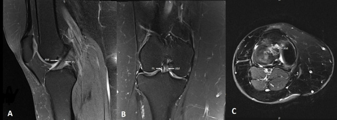
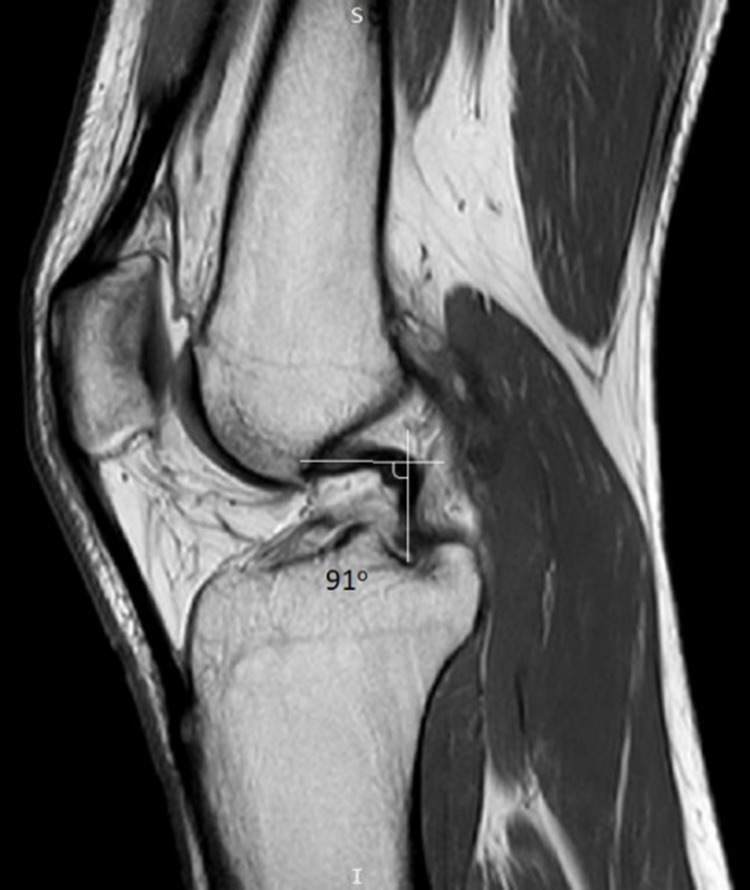
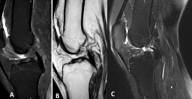
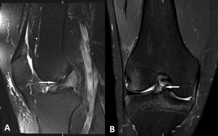
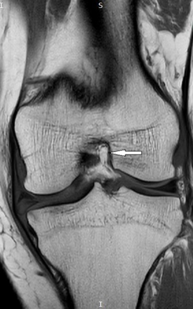
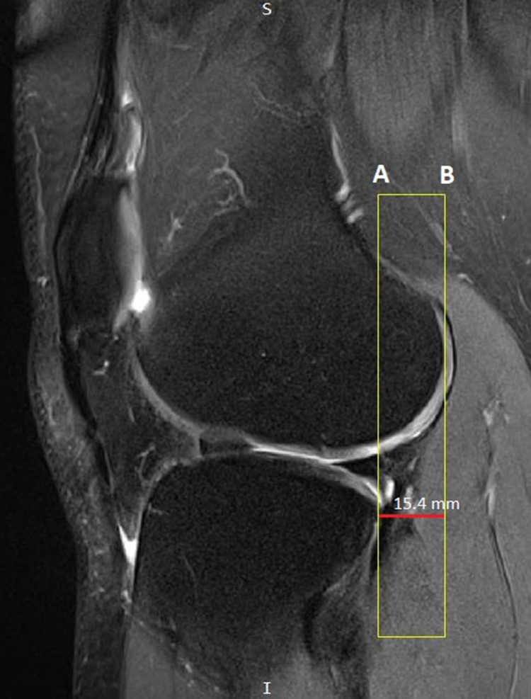
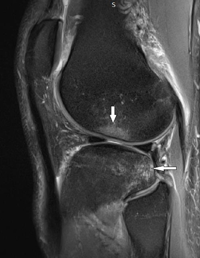
The Anterior Cruciate Ligament (ACL) is considered the most commonly injured ligament of the knee. Magnetic Resonance Imaging (MRI) has become an indispensable imaging tool and modality of choice for assessing and managing knee conditions. This article outlines an effective MRI imaging technique that improves the diagnostic accuracy of ACL injuries of the knee, covering the appropriate pulse sequences and optimal imaging planes. Furthermore, the article provides a comprehensive review of the appearance of complete and partial ACL tears on MRI in the acute and chronic phases. In addition, it identifies and illustrates the primary MRI signs (signs related to the absence or abnormal visualization of the ACL fibers) and secondary MRI signs (signs related to the mechanism of injury or associated knee injuries) of ACL injury.
前交叉韧带(ACL)被认为是膝关节最常受伤的韧带。磁共振成像(MRI)已成为评估和处理膝关节疾病不可或缺的成像工具和首选方式。本文概述了一种有效的MRI成像技术,该技术可提高膝关节ACL损伤的诊断准确性,涵盖了合适的脉冲序列和最佳成像平面。此外,本文还全面综述了急性和慢性期MRI上完全和部分ACL撕裂的表现。此外,它识别并说明了ACL损伤的主要MRI征象(与ACL纤维缺失或异常显影相关的征象)和次要MRI征象(与损伤机制或相关膝关节损伤相关的征象)。