The State Key Laboratory of Ophthalmology, Optometry and Visual Science, Wenzhou Medical University, Wenzhou, China.
Laboratory of Retinal Physiology and Disease, Eye Hospital and School of Ophthalmology and Optometry, Wenzhou Medical University, Wenzhou, China.
Invest Ophthalmol Vis Sci. 2024 Oct 1;65(12):19. doi: 10.1167/iovs.65.12.19.
Microglia-like cells derived from stem cells (iMG) provide a plentiful cell source for studying the functions of microglia in both normal and pathological conditions. Our goal is to establish a simplified and effective method for generating iMG in a precisely defined system. Additionally, we aim to achieve functional maturation of iMG through coculture with retinal organoids.
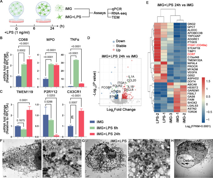
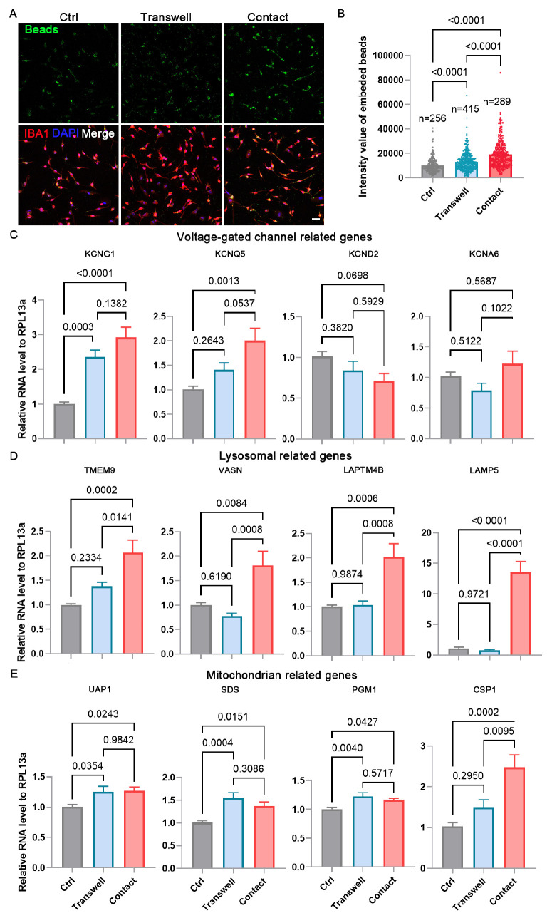
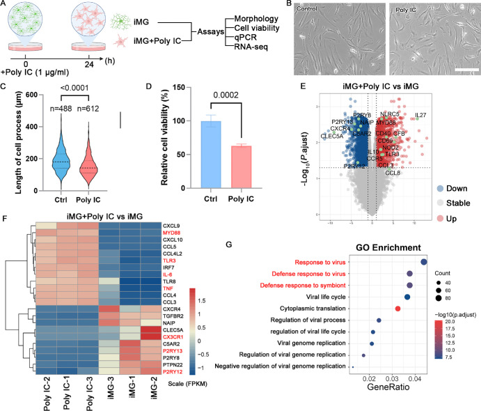
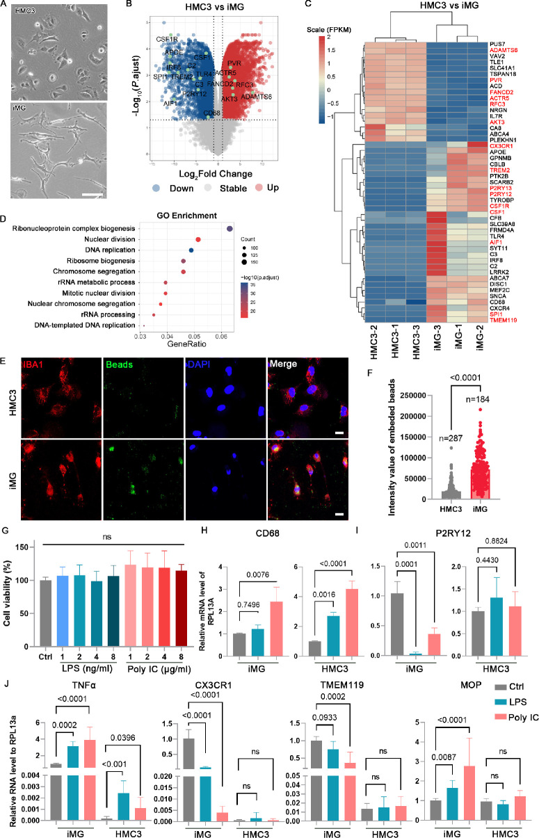
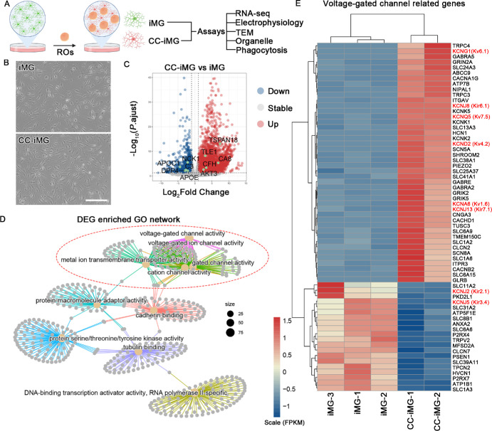
In this study, iMG were produced under precisely defined conditions. They were subjected to LPS and poly IC stimulation. Additionally, we examined distinct phenotypic and functional variances between iMG and HMC3, a commonly used human microglia cell line. To investigate how the retinal cell interaction enhances microglial properties, iMG were cocultured with retinal organoids, producing CC-iMG. We performed RNA sequencing, electrophysiological analysis, and transmission electron microscope (TEM) to examine the maturation of CC-iMG compared to iMG.
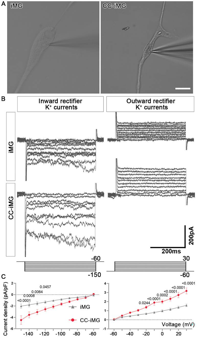
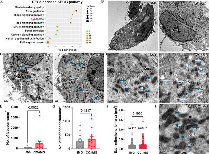
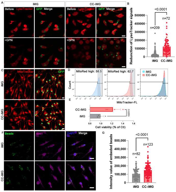
Our results demonstrated that iMG performed immune-responsive profiles closely resembling those of primary human microglia. Compared to HMC3, iMG expressed a higher level of typical microglial markers and exhibited enhanced phagocytic activity. The transcriptomic analysis uncovered notable alterations in the ion channel profile of CC-iMG compared to iMG. Electrophysiological examination demonstrated a heightened intensity of inward- and outward-rectifying K+ currents in CC-iMG. Furthermore, CC-iMG displayed elevated numbers of lysosomes and mitochondria, coupled with increased phagocytic activity.
These findings contribute to advancing our understanding of human microglial biology, specifically in characterizing and elucidating the functions of CC-iMG, thereby offering an in vitro microglial model for future scientific research and potential clinical applications in cell therapy.
源自干细胞的类小胶质细胞(iMG)为研究正常和病理条件下小胶质细胞的功能提供了丰富的细胞来源。我们的目标是在一个精确定义的系统中建立一种生成 iMG 的简化和有效的方法。此外,我们旨在通过与视网膜类器官共培养实现 iMG 的功能成熟。
在这项研究中,在精确定义的条件下产生 iMG。对其进行 LPS 和 poly IC 刺激。此外,我们还研究了 iMG 和 HMC3(一种常用的人小胶质细胞系)之间在表型和功能上的明显差异。为了研究视网膜细胞相互作用如何增强小胶质细胞特性,我们将 iMG 与视网膜类器官共培养,生成 CC-iMG。我们进行了 RNA 测序、电生理分析和透射电子显微镜(TEM)检查,以比较 CC-iMG 与 iMG 的成熟情况。
我们的结果表明,iMG 表现出与原代人小胶质细胞非常相似的免疫反应特征。与 HMC3 相比,iMG 表达了更高水平的典型小胶质细胞标志物,并表现出增强的吞噬活性。转录组分析揭示了 CC-iMG 与 iMG 相比离子通道谱的显著改变。电生理检查显示 CC-iMG 内向和外向整流 K+电流强度增加。此外,CC-iMG 显示出溶酶体和线粒体数量增加,同时吞噬活性增强。
这些发现有助于我们深入了解人类小胶质细胞生物学,特别是在表征和阐明 CC-iMG 的功能方面,从而为未来的科学研究和细胞治疗的潜在临床应用提供体外小胶质细胞模型。