Banerjee Atoshi, Lu Yimei, Do Kenny, Mize Travis, Wu Xiaogang, Chen Xiangning, Chen Jingchun
Nevada Institute of Personalized Medicine, University of Nevada, Las Vegas, NV, United States.
Department of Psychology, Nevada Institute of Personalized Medicine, University of Nevada, Las Vegas, NV, United States.
Front Cell Neurosci. 2021 Apr 9;15:629279. doi: 10.3389/fncel.2021.629279. eCollection 2021.
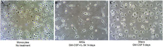
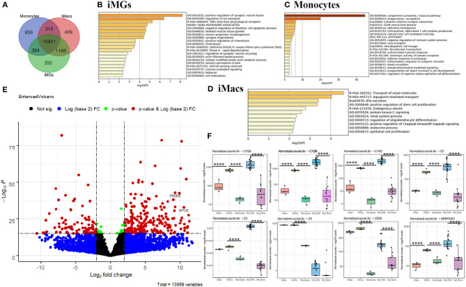
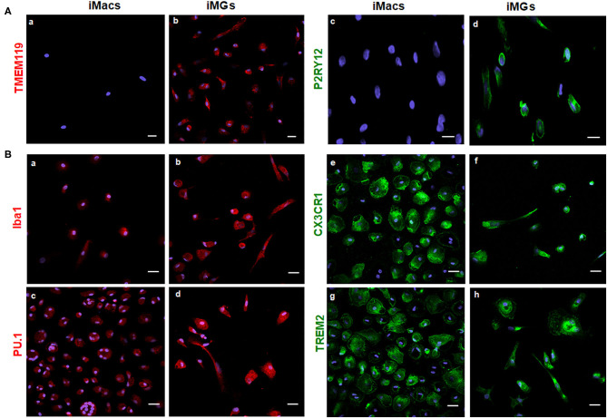
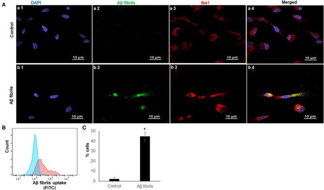
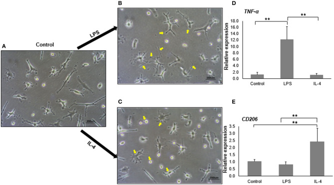
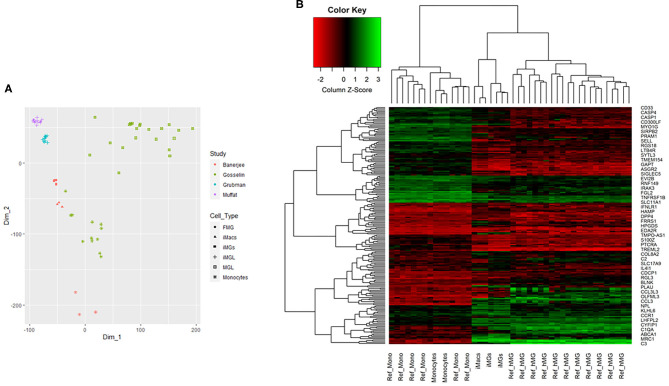
Microglia are the primary resident immune cells of the central nervous system that maintain physiological homeostasis in the brain and contribute to the pathogenesis of many psychiatric disorders and neurodegenerative diseases. Due to the lack of appropriate human cellular models, it is difficult to study the basic pathophysiological processes linking microglia to brain diseases. In this study, we adopted a microglia-like cellular model derived from peripheral blood monocytes with granulocyte-macrophage colony-stimulating factor (GM-CSF) and interleukin-34 (IL-34). We characterized and validated this cellular model by morphology, immunocytochemistry, gene expression profiles, and functional study. Our results indicated that the iMG cells developed typical microglial ramified morphology, expressed microglial specific surface markers (P2RY12 and TMEM119), and possessed phagocytic activity. Principal component analyses and multidimensional scaling analyses of RNA-seq data showed that iMG cells were distinct from monocytes and induced macrophages (iMacs) but clustered closer to human microglia and hiPSC-induced microglia. Heatmap analyses also found that iMG cells, but not monocytes, were closely clustered with human primary microglia. Further pathway and relative expression analysis indicated that unique genes from iMG cells were involved in the regulation of the complement system, especially in the synapse and ion transport. Overall, our data demonstrated that the iMG model mimicked many features of the brain resident microglia, highlighting its utility in the study of microglial function in many brain diseases, such as schizophrenia and Alzheimer's disease (AD).
小胶质细胞是中枢神经系统的主要常驻免疫细胞,它们维持大脑的生理稳态,并在许多精神疾病和神经退行性疾病的发病机制中发挥作用。由于缺乏合适的人类细胞模型,很难研究将小胶质细胞与脑部疾病联系起来的基本病理生理过程。在本研究中,我们采用了一种由外周血单核细胞与粒细胞-巨噬细胞集落刺激因子(GM-CSF)和白细胞介素-34(IL-34)衍生而来的小胶质细胞样细胞模型。我们通过形态学、免疫细胞化学、基因表达谱和功能研究对该细胞模型进行了表征和验证。我们的结果表明,诱导性小胶质细胞(iMG)形成了典型的小胶质细胞分支形态,表达小胶质细胞特异性表面标志物(P2RY12和TMEM119),并具有吞噬活性。RNA测序数据的主成分分析和多维尺度分析表明,iMG细胞与单核细胞和诱导性巨噬细胞(iMacs)不同,但与人类小胶质细胞和人诱导多能干细胞(hiPSC)诱导的小胶质细胞聚类更接近。热图分析还发现,iMG细胞而非单核细胞与人原代小胶质细胞紧密聚类。进一步的通路和相对表达分析表明,iMG细胞的独特基因参与补体系统的调节,尤其是在突触和离子转运方面。总体而言,我们的数据表明,iMG模型模拟了脑内常驻小胶质细胞的许多特征,突出了其在研究许多脑部疾病(如精神分裂症和阿尔茨海默病(AD))中小胶质细胞功能方面的实用性。