Wang Diyu, Lin Subin, Li Tuanwei, Yang Xiaohu, Zhong Xiang, Chen Qian, Jiang Guoqin, Li Chunyan
Department of General Surgery, Department of Orthopedics, The Second Affiliated Hospital of Soochow University, Suzhou, 215004, China.
CAS Key Laboratory of Nano-Bio Interface, Suzhou Key Laboratory of Functional Molecular Imaging Technology, Division of Nanobiomedicine and i-Lab, Suzhou Institute of Nano-Tech and Nano-Bionics, Chinese Academy of Sciences, Suzhou, 215123, China.
Mater Today Bio. 2024 Sep 26;29:101275. doi: 10.1016/j.mtbio.2024.101275. eCollection 2024 Dec.
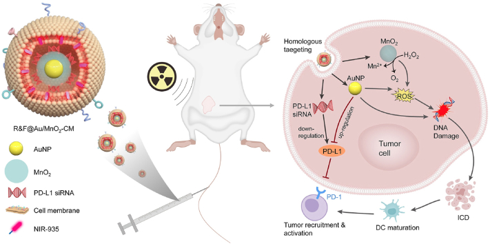
Radiotherapy plays a critical role in the clinical treatment of breast cancer. However, the efficacy of traditional X-ray radiotherapy is greatly limited by its low tumor specificity and treatment tolerance mediated by the tumor microenvironment. Herein, we proposed a novel nano-radiotherapy sensitization strategy to design and construct a cancer cell membrane-coated siRNA-decorated Au/MnO nanosensitizer (R&F@Au/MnO-CM) to synergistically enhance radio-immunotherapy for breast cancer. In the integrated nanosensitizer, the cancer cell membrane (CM) derived from 4T1 breast cancer cells is utilized for targeted functionality, while Au/MnO is designed to improve X-ray absorption and alleviate tumor hypoxia. Additionally, PD-L1 siRNA (R) is used to downregulate PD-L1 expression in tumor cells. In an in situ mouse model of 4T1 breast cancer, R&F@Au/MnO-CM demonstrated accurate tumor identification via CM-mediated homologous targeting after intravenous injection, which was monitored in real-time through NIR-II fluorescence imaging of NIR-935 (F). Subsequently, the radiotherapy sensitivity was achieved due to the strong radiation absorption properties and oxygen generation through catalysis of Au/MnO upon X-ray irradiation. Furthermore, the immunosuppressive microenvironment of the tumor is improved by downregulating PD-L1, enhancing synergistic anti-tumor effect. Our findings demonstrate a promising approach for tumor treatment by combining targeted enhanced radiotherapy with immune activation.
放射治疗在乳腺癌的临床治疗中起着关键作用。然而,传统的X射线放射治疗的疗效受到其低肿瘤特异性以及肿瘤微环境介导的治疗耐受性的极大限制。在此,我们提出了一种新型的纳米放射治疗增敏策略,设计并构建了一种癌细胞膜包裹的、用小干扰RNA(siRNA)修饰的金/二氧化锰纳米增敏剂(R&F@Au/MnO-CM),以协同增强乳腺癌的放射免疫治疗效果。在这种集成纳米增敏剂中,源自4T1乳腺癌细胞的癌细胞膜(CM)用于实现靶向功能,而金/二氧化锰的设计目的是提高X射线吸收并缓解肿瘤缺氧。此外,PD-L1 siRNA(R)用于下调肿瘤细胞中PD-L1的表达。在4T1乳腺癌原位小鼠模型中,R&F@Au/MnO-CM在静脉注射后通过CM介导的同源靶向作用表现出准确的肿瘤识别能力,这通过NIR-935的近红外二区(NIR-II)荧光成像(F)进行实时监测。随后,由于金/二氧化锰在X射线照射下具有强烈的辐射吸收特性并能催化产生氧气,从而实现了放射治疗敏感性的提高。此外,通过下调PD-L1改善了肿瘤的免疫抑制微环境,增强了协同抗肿瘤效果。我们的研究结果表明,将靶向增强放射治疗与免疫激活相结合是一种很有前景的肿瘤治疗方法。