Kanoda Ryo, Kikuchi Tomohiro, Utsumi Akihito, Mochizuki Shotaro, Matsuishi Akira, Kaneta Akinao, Nirei Azuma, Hanayama Hiroyuki, Saze Zenichiro, Hikichi Takuto, Hashimoto Yuko, Kono Koji
Department of Gastrointestinal Tract Surgery, Fukushima Medical University School of Medicine, 1 Hikariga-Oka, Fukushima City, Fukushima, 960-1295, Japan.
Department of Diagnostic Pathology, Fukushima Medical University School of Medicine, 1 Hikariga-Oka, Fukushima City, Fukushima, Japan.
Surg Case Rep. 2024 Oct 18;10(1):237. doi: 10.1186/s40792-024-02045-y.
Esophageal gastrointestinal stromal tumors (GISTs) are relatively rare, accounting for 2-5% of all GISTs. Typically, the treatment is surgery in nature. However, no standard procedure established for esophageal GISTs, and in many cases, subtotal esophagectomy or local resection via thoracoscopy or mediastinoscopy is performed. Thoracoscopic and endoscopic cooperative surgery (TECS) is a surgical approach similar to laparoscopic and endoscopic cooperative surgery used for gastric GIST; however, no reports of its use for esophageal GIST have been published to date. We herein report such a case along with a review of past literature.
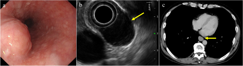
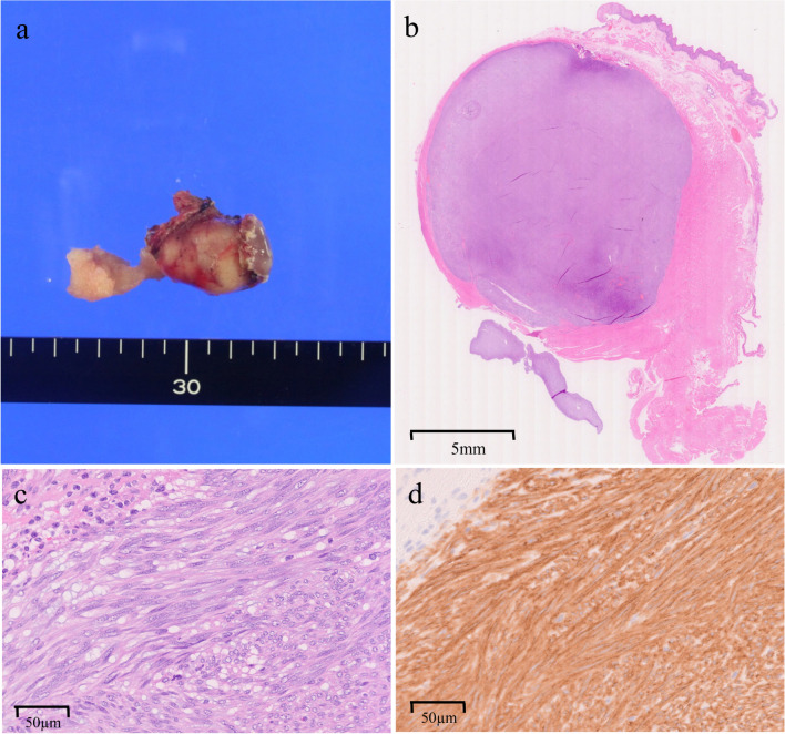
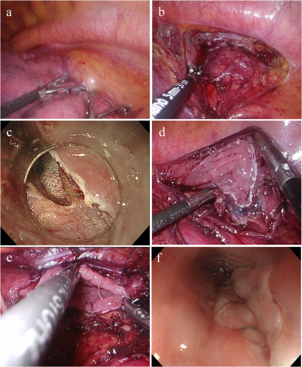
The patient was a 60-year-old man. Upper gastrointestinal contrast imaging revealed a subepithelial lesion in the esophagus. An 18 × 17 mm subepithelial lesion was identified in the left wall, 35 cm from the upper incisors, during upper gastrointestinal endoscopy, and was diagnosed as a GIST through endoscopic ultrasound-guided fine needle biopsy. TECS was therefore performed. The patient was placed in a prone position with his face to the left. After confirming the lesion under endoscopy and left thoracoscopy, the periesophageal area of the lesion was dissected under thoracoscopy. Subsequently, an endoscopic full-layer resection was performed. Finally, the excision site of the lesion was sutured under thoracoscopy. The operation took a total of 3 h and 22 min, with a blood loss of 50 mL.
The appropriate surgical procedure for esophageal GIST should be considered according to the location and size of the lesion. TECS ensures that the resection margins are secured using an endoscopic or thoracoscopic approach. Furthermore, TECS is minimally invasive, avoiding esophagectomy and reconstruction, which makes it a potential surgical option for esophageal GISTs.
食管胃肠道间质瘤(GISTs)相对罕见,占所有GISTs的2%-5%。通常,其治疗本质上是手术治疗。然而,目前尚未建立针对食管GISTs的标准手术程序,在许多情况下,会进行食管次全切除术或通过胸腔镜或纵隔镜进行局部切除术。胸腔镜与内镜联合手术(TECS)是一种类似于用于胃GIST的腹腔镜与内镜联合手术的手术方法;然而,迄今为止尚无其用于食管GIST的报道。我们在此报告这样一例病例并回顾既往文献。
患者为一名60岁男性。上消化道造影显示食管有一个上皮下病变。上消化道内镜检查时,在距上切牙35cm处的左壁发现一个18×17mm的上皮下病变,经内镜超声引导下细针穿刺活检诊断为GIST。因此进行了TECS手术。患者俯卧位,面部朝左。在内镜和左胸腔镜下确认病变后,在胸腔镜下对病变周围的食管区域进行解剖。随后,进行了内镜全层切除术。最后,在胸腔镜下对病变切除部位进行缝合。手术共耗时3小时22分钟,失血50mL。
应根据病变的位置和大小考虑适合食管GIST的手术程序。TECS可通过内镜或胸腔镜方法确保切缘安全。此外,TECS微创,避免了食管切除术和重建术,这使其成为食管GIST的一种潜在手术选择。