Theodor Kocher Institute, University of Bern, Freiestrasse 1, Bern, CH-3012, Switzerland.
Department of Chemistry, Biochemistry and Pharmaceutical Sciences, University of Bern, Bern, Switzerland.
J Neuroinflammation. 2024 Oct 23;21(1):272. doi: 10.1186/s12974-024-03247-9.
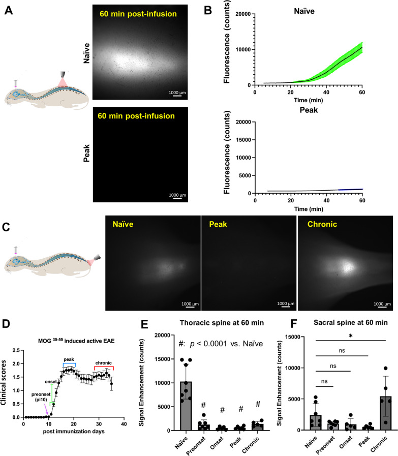
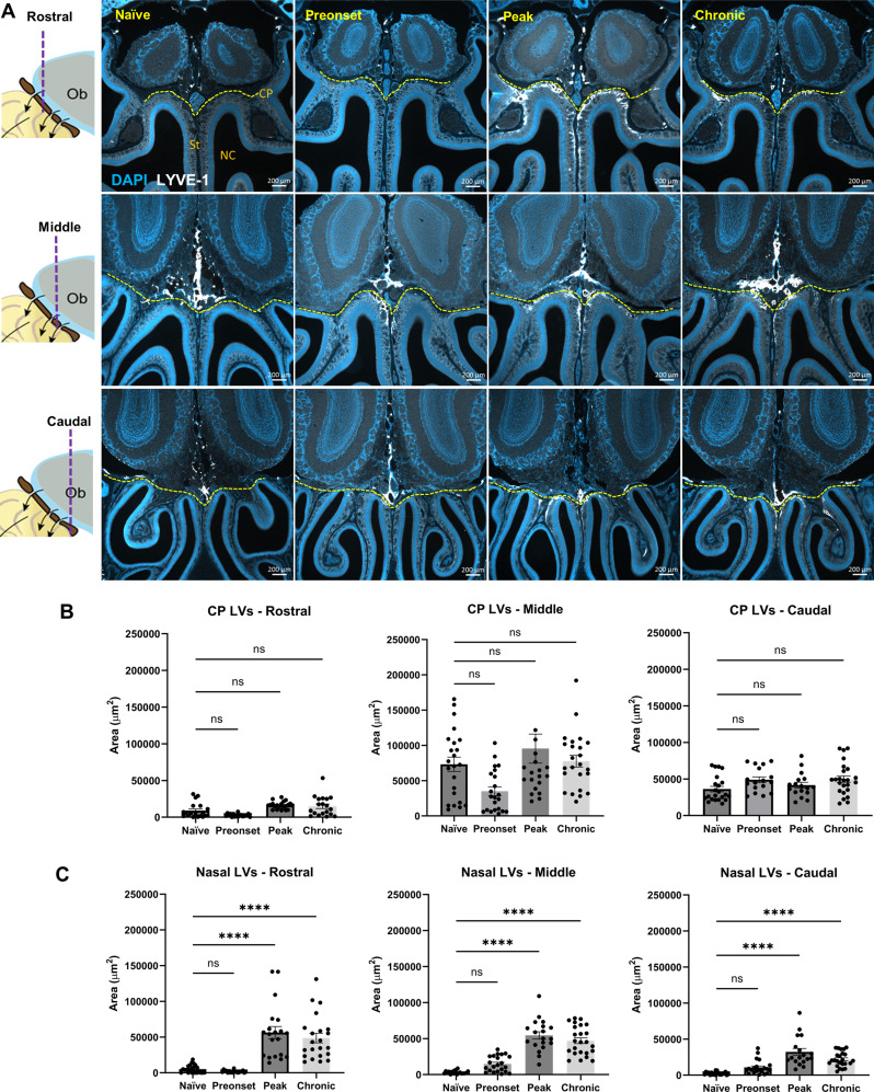
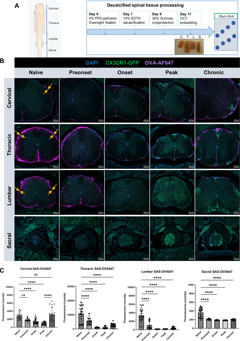
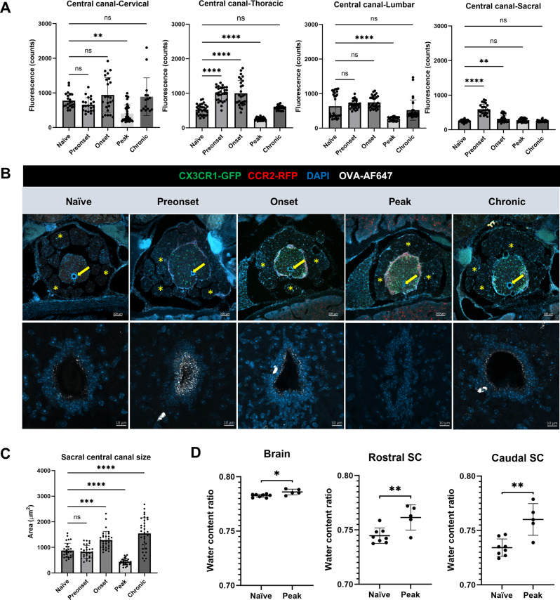
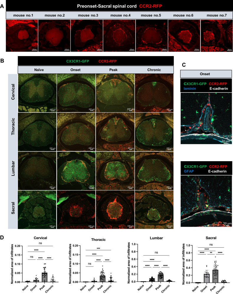
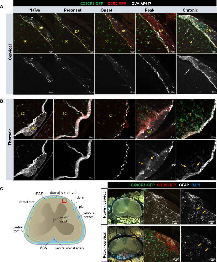
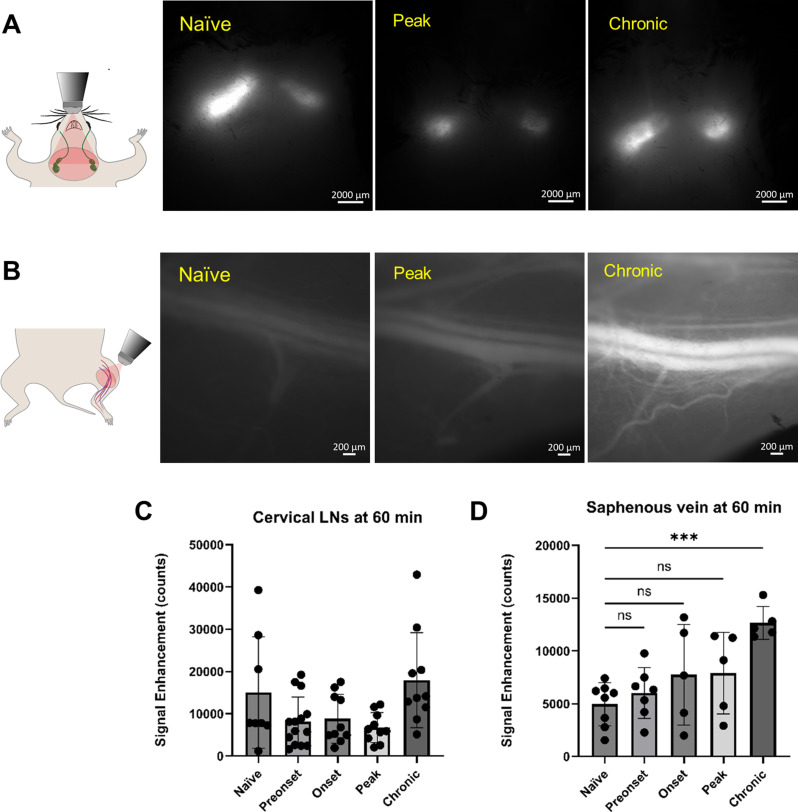
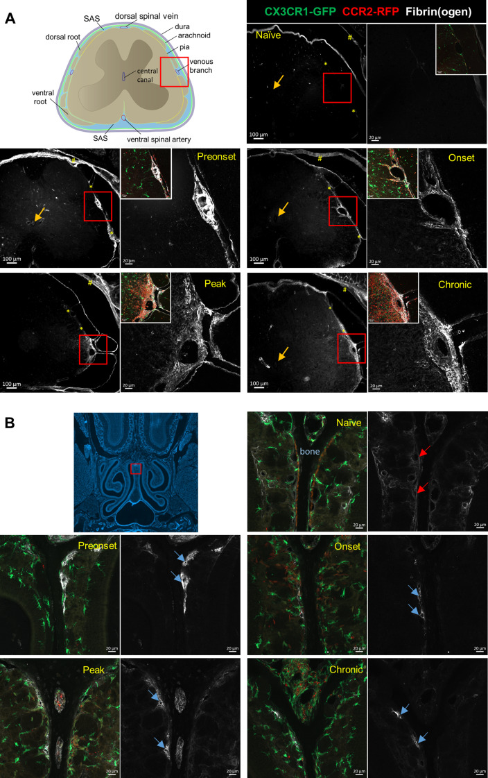
Accumulation of immune cells and proteins in the subarachnoid space (SAS) is found during multiple sclerosis and in the animal model experimental autoimmune encephalomyelitis (EAE). Whether the flow of cerebrospinal fluid (CSF) along the SAS of the spinal cord is impacted is yet unknown. Combining intravital near-infrared (NIR) imaging with histopathological analyses, we observed a significantly impaired bulk flow of CSF tracers within the SAS of the spinal cord prior to EAE onset, which persisted until peak stage and was only partially recovered during chronic disease. The impairment of spinal CSF flow coincided with the appearance of fibrin aggregates in the SAS, however, it preceded immune cell infiltration and breakdown of the glia limitans superficialis. Conversely, cranial CSF efflux to cervical lymph nodes was not altered during the disease course. Our study highlights an early and persistent impairment of spinal CSF flow and suggests it as a sensitive imaging biomarker for pathological changes within the leptomeninges.
在多发性硬化症和实验性自身免疫性脑脊髓炎(EAE)动物模型中,蛛网膜下腔(SAS)中免疫细胞和蛋白质的积累。目前尚不清楚脑脊液(CSF)沿脊髓 SAS 的流动是否受到影响。通过将活体近红外(NIR)成像与组织病理学分析相结合,我们在 EAE 发作前观察到脊髓 SAS 内 CSF 示踪剂的整体流动明显受损,这种情况一直持续到高峰期,并在慢性疾病期间仅部分恢复。脊髓 CSF 流动的受损与 SAS 中纤维蛋白聚集体的出现相吻合,但它先于免疫细胞浸润和软脑膜表面胶质界限的破坏。相反,在疾病过程中,颅 CSF 流出到颈部淋巴结没有改变。我们的研究强调了脊髓 CSF 流动的早期和持续受损,并将其作为脑膜内病理变化的敏感成像生物标志物。