Jánosi Kinga Mária, Cerghizan Diana, Bai Eszter Elza, Mureșan Izabella Éva, Kovács Alpár, Szász Andrea, Hulpe Adrian, Markovics Emese Rita, Mártha Krisztina Ildikó, Pop Silvia Izabella
Faculty of Dental Medicine, George Emil Palade University of Medicine, Pharmacy, Science and Technology of Targu Mureș, 38 Gh. Marinescu Str., 540139 Targu Mureș, Romania.
Private Practice, 540501 Targu Mureș, Romania.
Dent J (Basel). 2024 Sep 29;12(10):313. doi: 10.3390/dj12100313.
Intraoral scanning technology has opened new perspectives in dental practice, and combined with CAD/CAM technology, contributes significantly to fabricating high-quality prosthetic restorations. Our in vitro study aims to assess the accuracy of digital models obtained from one laboratory and two less commonly used intraoral scanners by conducting 3D measurements on the digital models obtained.
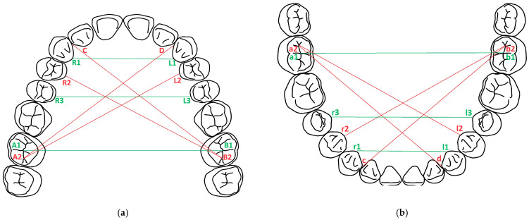
An articulated simulator cast was used. Forty-eight scans were performed before and after tooth preparation with each scanner. The Zeiss Inspect software (Version: 2023.3.0.969) was used for measurements in sagittal and transversal planes. The obtained values were compared to reference values resulting from manual measurements.
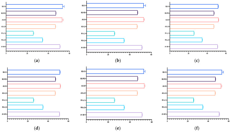
Digital impressions provided discrepancies compared to the reference model. The lowest differences at the A2-L2 (the diagonal dimension of the models from the distal fossa of the second right maxillary molar and the maximum oral convexity of the artificial gingiva at the first left premolar) and the A1-B1 (transversal dimension of the model in the posterior area, from the right second molar's occlusal central fossa to the left second molar central fossa) distances were obtained for the upper models, and at the a1-b1 distance for all the lower models, except the non-prepared models scanned with the intraoral scanners (the discrepancies were not statistically significant). The discrepancies increased with the distance from the starting point of the scan.
The number and position of prepared teeth can influence the accuracy of the scans. Distortions can appear in the case of multiple preparations. The scanning protocol and calibration must be optimized for the highest accuracy. Furthermore, in vivo studies are necessary to evaluate the clinical applicability of these findings.
口腔内扫描技术为牙科实践开辟了新视野,与计算机辅助设计/计算机辅助制造(CAD/CAM)技术相结合,对制作高质量的修复体有显著贡献。我们的体外研究旨在通过对所获得的数字模型进行三维测量,评估从一个实验室和两台较少使用的口腔内扫描仪获得的数字模型的准确性。
使用了一个关节式模拟模型。每种扫描仪在牙齿预备前后各进行48次扫描。使用蔡司Inspect软件(版本:2023.3.0.969)在矢状面和横断面进行测量。将获得的值与手动测量得到的参考值进行比较。
与参考模型相比,数字印模存在差异。在上颌模型中,在A2-L2(从右上颌第二磨牙远中窝到左第一前磨牙处人工牙龈最大口腔凸度的模型对角线尺寸)和A1-B1(模型后部区域的横向尺寸,从右第二磨牙咬合中央窝到左第二磨牙中央窝)距离处获得的差异最小;在下颌所有模型中,除了用口腔内扫描仪扫描的未预备模型外,在a1-b1距离处差异最小(差异无统计学意义)。差异随着距扫描起点的距离增加而增大。
预备牙齿的数量和位置会影响扫描的准确性。在多次预备的情况下可能会出现变形。必须优化扫描方案和校准以获得最高精度。此外,有必要进行体内研究以评估这些发现的临床适用性。