Krüger-Genge Anne, Harb Kudor, Braune Steffen, Jung Conrad H G, Westphal Sophia, Bär Stefanie, Mauger Olivia, Küpper Jan-Heiner, Jung Friedrich
Life Science and Bioprocesses, Fraunhofer Institute for Applied Polymer Research (IAP), 14476 Potsdam, Germany.
Institute of Biotechnology, Molecular Cell Biology, Brandenburg University of Technology Cottbus-Senftenberg, 01968 Senftenberg, Germany.
Life (Basel). 2024 Oct 1;14(10):1253. doi: 10.3390/life14101253.
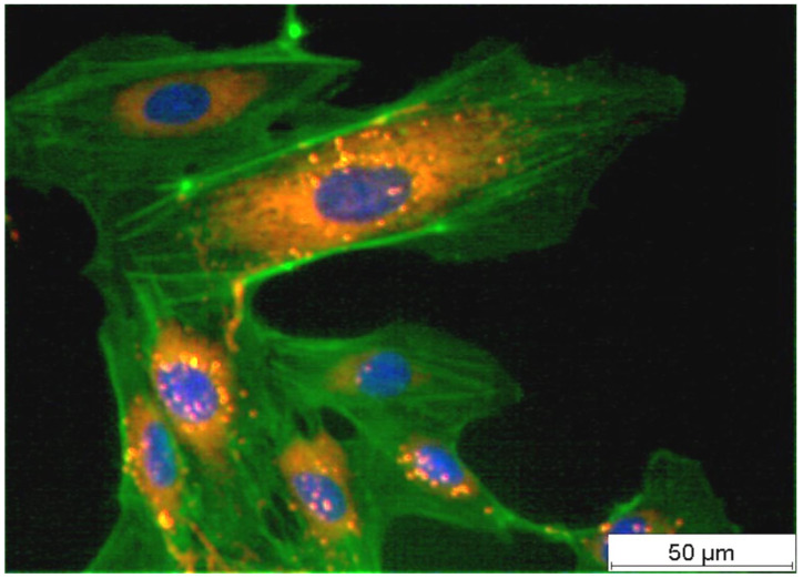
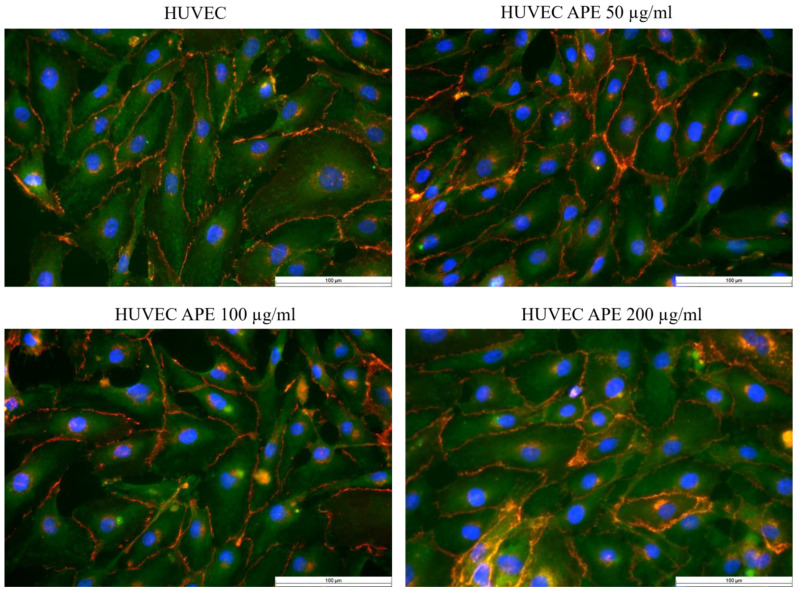
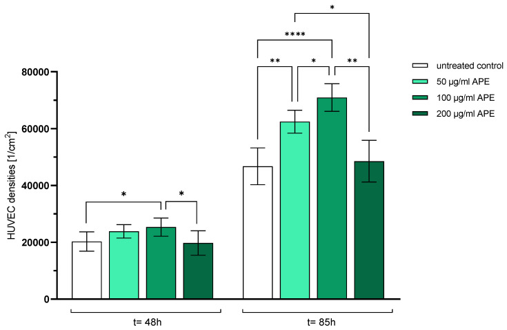
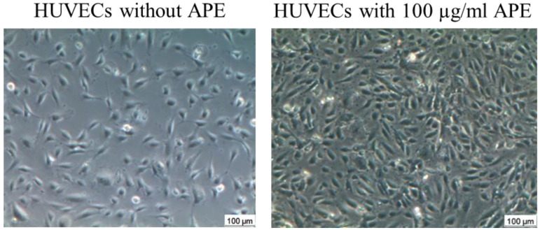
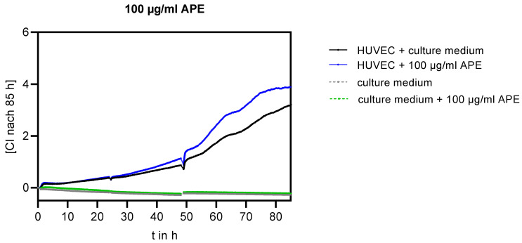
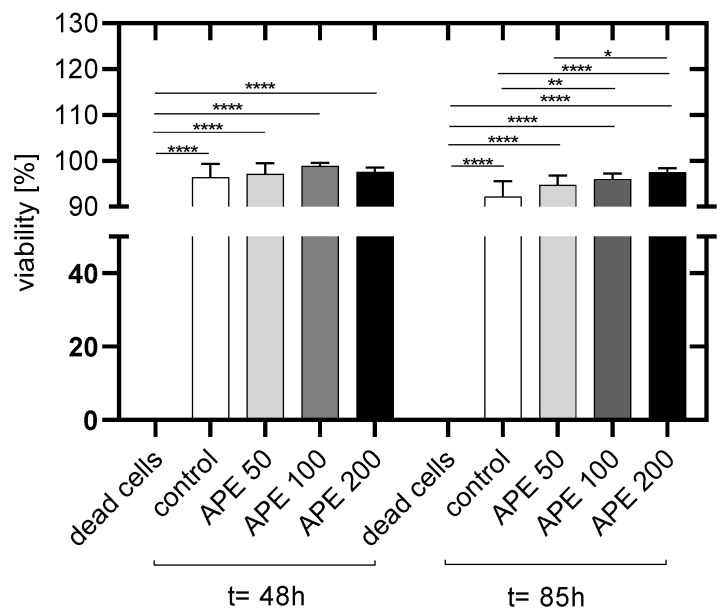
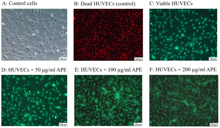
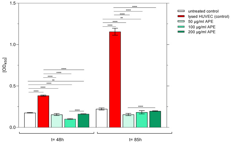
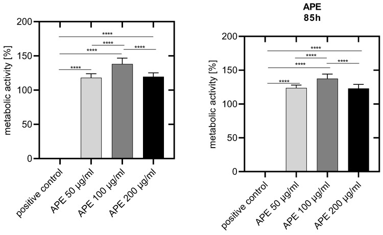
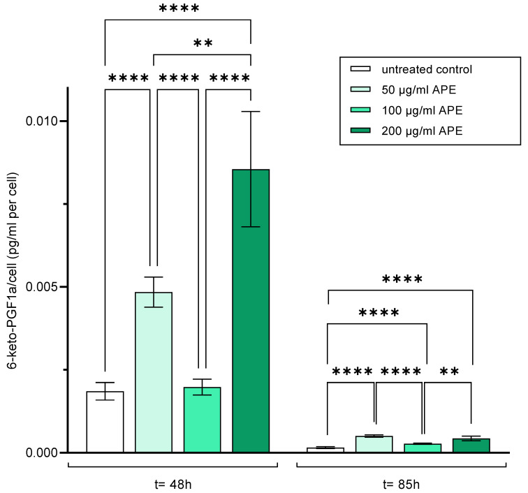
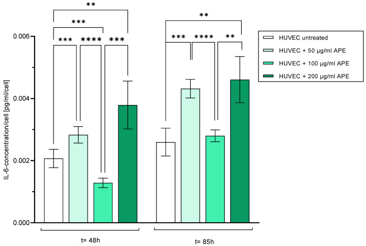
Atherosclerosis is initiated by injury or damage to the vascular endothelial cell monolayer. Therefore, the early repair of the damaged vascular endothelium by a proliferation of neighbouring endothelial cells is important to prevent atherosclerosis and thrombotic events. (AP) has been used as a dietary supplement, mainly due to its high content of vitamins, minerals, amino acids, and pigments such as chlorophylls, carotenoids, and phycocyanin, ingredients with antioxidant, anti-inflammatory, and anti-thrombotic properties. Therefore, in this prospective, placebo-controlled, data-driven, sample-size-estimated in vitro study, we tested whether an aqueous extract of AP at different concentrations (50, 100, and 200 µg/mL) had an effect on the different cellular parameters of human umbilical vein endothelial cells. Therefore, cell impedance measurement and cell proliferation were measured to investigate the monolayer formation. In addition, cell viability, integrity, and metabolism were analysed to evaluate singular cellular functions, especially the antithrombotic state. Furthermore, cell-cell and cell-substrate interactions were observed. The highest proliferation was achieved after the addition of 100 µg/mL. This was consistently confirmed by two independent optical experiments in cell cultures 48 h and 85 h after seeding and additionally by an indirect test. At this concentration, the activation or dysfunction of HUVECs was completely prevented, as confirmed by prostacyclin and interleukin-6 levels. In conclusion, in this study, AP induced a significant increase in HUVEC proliferation without inducing an inflammatory response but altered the hemostasiological balance in favour of prostacyclin over thromboxane, thereby creating an antithrombotic state. Thus, APE could be applied in the future as an accelerator of endothelial cell proliferation after, e.g., stent placement or atherosclerosis.
动脉粥样硬化由血管内皮细胞单层的损伤引发。因此,相邻内皮细胞增殖对受损血管内皮进行早期修复,对于预防动脉粥样硬化和血栓形成事件至关重要。紫球藻(AP)一直被用作膳食补充剂,主要因其富含维生素、矿物质、氨基酸以及叶绿素、类胡萝卜素和藻蓝蛋白等色素,这些成分具有抗氧化、抗炎和抗血栓特性。因此,在这项前瞻性、安慰剂对照、数据驱动、样本量估算的体外研究中,我们测试了不同浓度(50、100和200 µg/mL)的AP水提取物对人脐静脉内皮细胞不同细胞参数的影响。因此,通过测量细胞阻抗和细胞增殖来研究单层形成。此外,分析细胞活力、完整性和代谢以评估单一细胞功能,尤其是抗血栓状态。此外,还观察了细胞 - 细胞和细胞 - 底物相互作用。添加100 µg/mL后增殖达到最高。这在接种后48小时和85小时的细胞培养中的两项独立光学实验以及另外一项间接测试中得到了一致证实。在此浓度下,如前列环素和白细胞介素 - 6水平所证实,人脐静脉内皮细胞(HUVECs)的激活或功能障碍被完全预防。总之,在本研究中,AP显著增加了HUVEC的增殖,且未引发炎症反应,但改变了止血平衡,有利于前列环素而非血栓素,从而形成抗血栓状态。因此,未来紫球藻提取物(APE)可作为例如支架置入或动脉粥样硬化后内皮细胞增殖的促进剂应用。