Department of Critical Care Medicine, Cliniques universitaires Saint Luc, Université catholique de Louvain (UCLouvain), Avenue Hippocrate 10, 1200, Brussels, Belgium.
Pôle de Pneumologie, O.R.L. et Dermatologie (LuNS, Lung-Nose-Skin), Institut de Recherche Expérimentale et Clinique, Université catholique de Louvain (UCLouvain), Brussels, Belgium.
Crit Care. 2024 Oct 30;28(1):350. doi: 10.1186/s13054-024-05127-3.
The airway epithelium (AE) fulfils multiple functions to maintain pulmonary homeostasis, among which ensuring adequate barrier function, cell differentiation and polarization, and actively transporting immunoglobulin A (IgA), the predominant mucosal immunoglobulin in the airway lumen, via the polymeric immunoglobulin receptor (pIgR). Morphological changes of the airways have been reported in ARDS, while their detailed features, impact for mucosal immunity, and causative mechanisms remain unclear. Therefore, this study aimed to assess epithelial alterations in the distal airways of patients with ARDS.
We retrospectively analyzed lung tissue samples from ARDS patients and controls to investigate and quantify structural and functional changes in the small airways, using multiplex fluorescence immunostaining and computer-assisted quantification on whole tissue sections. Additionally, we measured markers of mucosal immunity, IgA and pIgR, alongside with other epithelial markers, in the serum and the broncho-alveolar lavage fluid (BALF) prospectively collected from ARDS patients and controls.
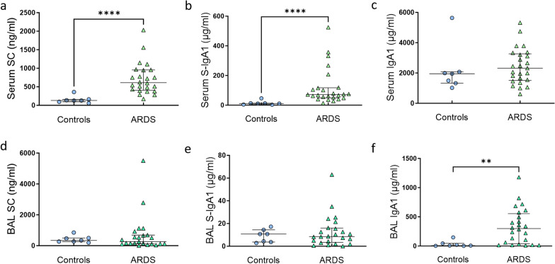
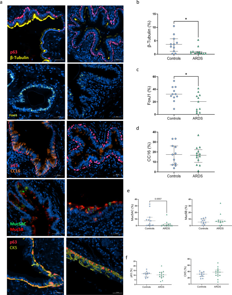
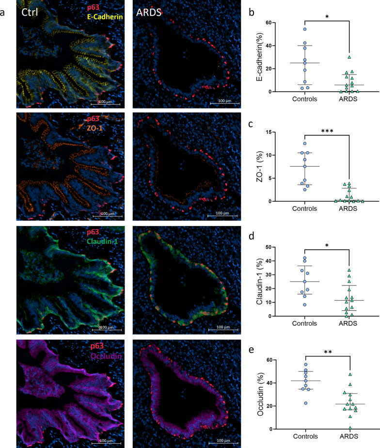
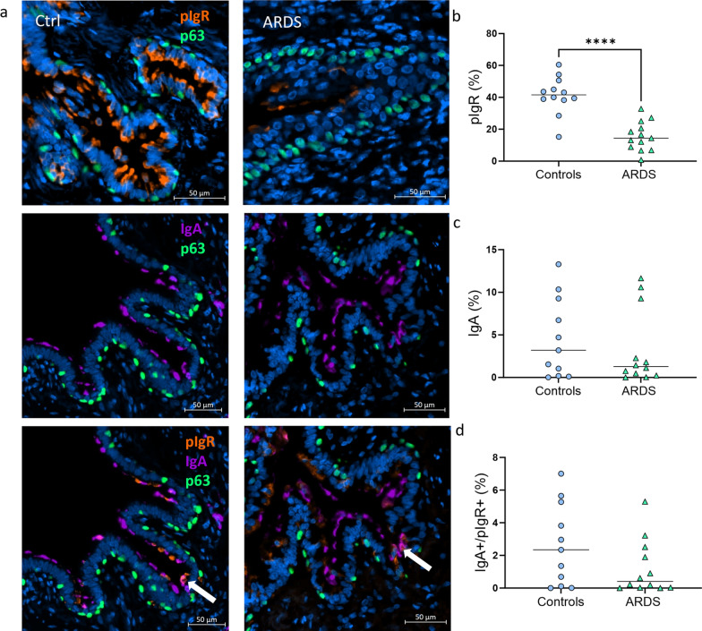
Compared to controls, airways of ARDS were characterized by increased epithelial denudation (p = 0.0003) and diffuse epithelial infiltration by neutrophils (p = 0.0005). Quantitative evaluation of multiplex fluorescence immunostaining revealed a loss of ciliated cells (p = 0.0317) a trend towards decreased goblet cells (p = 0.056), and no change regarding cell progenitors (basal and club cells), indicating altered mucociliary differentiation. Increased epithelial permeability was also shown in ARDS with a significant decrease of tight (p < 0.0001) and adherens (p = 0.025) junctional proteins. Additionally, we observed a significant decrease of the expression of pIgR, (p < 0.0001), indicating impaired mucosal IgA immunity. Serum concentrations of secretory component (SC) and S-IgA were increased in ARDS (both p < 0.0001), along other lung-derived proteins (CC16, SP-D, sRAGE). However, their BALF concentrations remained unchanged, suggesting a spillover of airway and alveolar proteins through a damaged AE.
The airway epithelium from patients with ARDS exhibits multifaceted alterations leading to altered mucociliary differentiation, compromised defense functions and increased permeability with pneumoproteinemia.
气道上皮 (AE) 具有多种功能,以维持肺内环境稳定,其中包括确保充分的屏障功能、细胞分化和极化,并通过多免疫球蛋白受体 (pIgR) 主动转运气道腔中的主要黏膜免疫球蛋白 IgA。在 ARDS 中已经报道了气道的形态变化,但其详细特征、对黏膜免疫的影响和因果机制尚不清楚。因此,本研究旨在评估 ARDS 患者远端气道的上皮变化。
我们回顾性分析了 ARDS 患者和对照组的肺组织样本,使用多色荧光免疫染色和全组织切片的计算机辅助定量分析来研究和量化小气道的结构和功能变化。此外,我们前瞻性地从 ARDS 患者和对照组中收集血清和支气管肺泡灌洗液 (BALF) 中测量黏膜免疫标志物 IgA 和 pIgR 以及其他上皮标志物。
与对照组相比,ARDS 患者的气道表现为上皮脱落增加(p=0.0003)和中性粒细胞弥漫性上皮浸润(p=0.0005)。多色荧光免疫染色的定量评估显示纤毛细胞丢失(p=0.0317),杯状细胞减少(p=0.056),细胞祖代(基底细胞和 club 细胞)无变化,表明黏液纤毛分化改变。上皮通透性增加也在 ARDS 中表现出来,紧密(p<0.0001)和黏附(p=0.025)连接蛋白显著减少。此外,我们观察到 pIgR 的表达显著降低(p<0.0001),表明黏膜 IgA 免疫受损。ARDS 患者的血清分泌成分 (SC) 和 S-IgA 浓度增加(均 p<0.0001),以及其他肺源性蛋白(CC16、SP-D、sRAGE)。然而,它们的 BALF 浓度保持不变,表明通过受损的 AE 发生了气道和肺泡蛋白的溢出。
ARDS 患者的气道上皮表现出多种改变,导致黏液纤毛分化改变、防御功能受损和通透性增加,并伴有肺蛋白血症。