• 文献检索
  • 文档翻译
  • 深度研究
  • 学术资讯
  • Suppr Zotero 插件Zotero 插件
  • 邀请有礼
  • 套餐&价格
  • 历史记录
应用&插件
Suppr Zotero 插件Zotero 插件浏览器插件Mac 客户端Windows 客户端微信小程序
定价
高级版会员购买积分包购买API积分包
服务
文献检索文档翻译深度研究API 文档MCP 服务
关于我们
关于 Suppr公司介绍联系我们用户协议隐私条款
关注我们

Suppr 超能文献

核心技术专利:CN118964589B侵权必究
粤ICP备2023148730 号-1Suppr @ 2026

文献检索

告别复杂PubMed语法,用中文像聊天一样搜索,搜遍4000万医学文献。AI智能推荐,让科研检索更轻松。

立即免费搜索

文件翻译

保留排版,准确专业,支持PDF/Word/PPT等文件格式,支持 12+语言互译。

免费翻译文档

深度研究

AI帮你快速写综述,25分钟生成高质量综述,智能提取关键信息,辅助科研写作。

立即免费体验

使用微创 PicoGreen 标记策略追踪小型细胞外囊泡。

Tracking Small Extracellular Vesicles Using a Minimally Invasive PicoGreen Labeling Strategy.

机构信息

Department of Pathology and Laboratory Medicine, University of Kansas Medical Center, Kansas City, Kansas 66160, United States.

Department of Cancer Biology, University of Kansas Medical Center, Kansas City, Kansas 66160, United States.

出版信息

ACS Appl Bio Mater. 2024 Nov 18;7(11):7770-7783. doi: 10.1021/acsabm.4c01500. Epub 2024 Oct 31.

DOI:10.1021/acsabm.4c01500
PMID:39482871
原文链接:https://pmc.ncbi.nlm.nih.gov/articles/PMC11577420/
Abstract

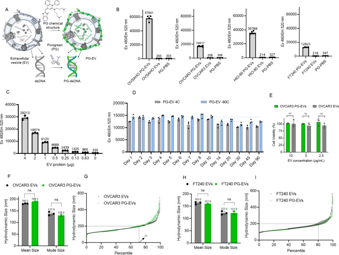
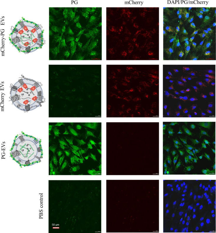
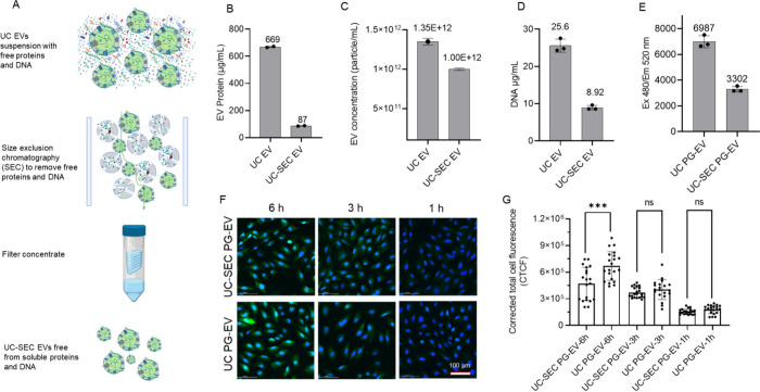
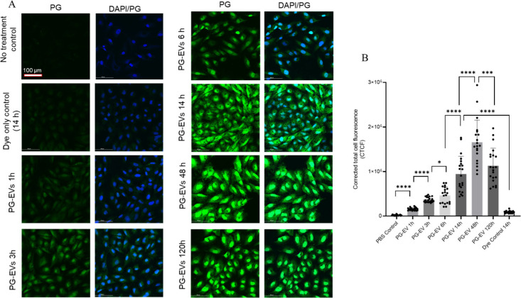
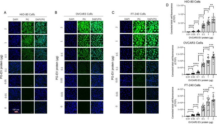
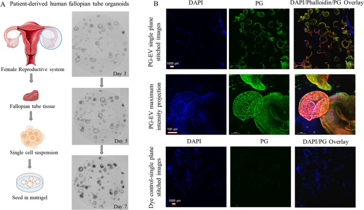
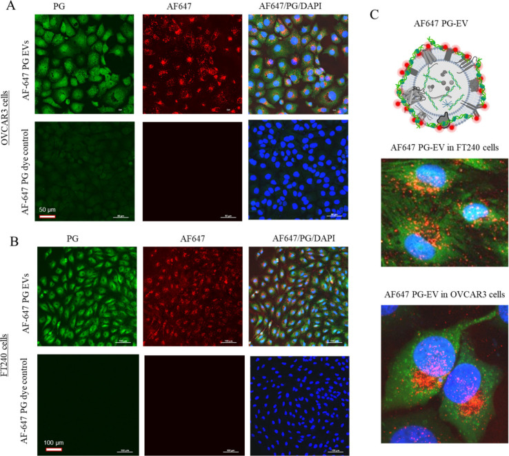
Extracellular vesicles (EVs) are cell-secreted lipid bilayer delimited particles that mediate cellular communication. These tiny sacs of cellular information play an important role in cell communication and alter the physiological process under both normal and pathological conditions. As such, tracking EVs can provide valuable information regarding the basic understanding of cell communication, the onset of early malignancy, and biomarker discovery. Most of the current EV-tracking strategies are invasive, altering the natural characteristics of EVs by modifying the lipid bilayer with lipophilic dyes or surface proteins with fluorescent reporters. The invasive labeling strategies could alter the natural processes of EVs and thereby have major limitations for functional studies. Here, we report an alternative minimally invasive EV labeling strategy using PicoGreen (PG), a small molecule that fluoresces at 520 nm when bound to dsDNA. We show that PG binds to dsDNA associated with small EVs (50-200 nm), forming a stable and highly fluorescent PG-DNA complex in EVs (PG-EVs). In both 2D cell culture and 3D organoid models, PG-EV showed efficient tracking properties, including a high signal-to-noise ratio, time- and concentration-dependent uptake, and the ability to traverse a 3D environment. We further validated PG-EV tracking using dual-labeled EVs following two orthogonal labeling strategies: (1) Bioconjugation via surface amine labeling and (2) donor cell engineering via endogenously expressing mCherry-tetraspanin (CD9/CD63/CD81) reporter proteins. Our study has shown the feasibility of using PG-EV as an effective EV tracking strategy that can be applied for studying the functional role of EVs across multiple model systems.

摘要

细胞外囊泡 (EVs) 是由细胞分泌的双层脂质膜限定的颗粒,介导细胞间通讯。这些微小的细胞信息囊泡在正常和病理条件下的细胞通讯和改变生理过程中发挥着重要作用。因此,跟踪 EVs 可以提供有关细胞通讯基本理解、早期恶性肿瘤发生和生物标志物发现的有价值信息。目前大多数 EV 追踪策略都是侵入性的,通过用亲脂性染料或荧光报告蛋白修饰脂质双层来改变 EV 的天然特征,从而对 EV 进行标记。这种侵入性的标记策略可能会改变 EV 的自然过程,因此对功能研究有很大的局限性。在这里,我们报告了一种使用 PicoGreen(PG)的替代微创 EV 标记策略,PG 是一种小分子,当与双链 DNA(dsDNA)结合时会在 520nm 处发出荧光。我们表明,PG 与与小 EV(50-200nm)相关的 dsDNA 结合,在 EV 中形成稳定且高度荧光的 PG-DNA 复合物(PG-EVs)。在 2D 细胞培养和 3D 类器官模型中,PG-EV 均表现出高效的追踪特性,包括高信噪比、时间和浓度依赖性摄取以及穿越 3D 环境的能力。我们进一步通过两种正交标记策略(1)通过表面胺标记进行生物共轭和(2)通过内源性表达 mCherry-四跨膜蛋白(CD9/CD63/CD81)报告蛋白进行供体细胞工程来验证 PG-EV 追踪。我们的研究表明,使用 PG-EV 作为一种有效的 EV 追踪策略是可行的,可以应用于在多个模型系统中研究 EV 的功能作用。

https://cdn.ncbi.nlm.nih.gov/pmc/blobs/edfe/11577420/e9c73cbbacde/mt4c01500_0007.jpg
https://cdn.ncbi.nlm.nih.gov/pmc/blobs/edfe/11577420/e4efcc3397af/mt4c01500_0001.jpg
https://cdn.ncbi.nlm.nih.gov/pmc/blobs/edfe/11577420/71b5d304fe9d/mt4c01500_0002.jpg
https://cdn.ncbi.nlm.nih.gov/pmc/blobs/edfe/11577420/fce3dc44000a/mt4c01500_0003.jpg
https://cdn.ncbi.nlm.nih.gov/pmc/blobs/edfe/11577420/4de5bf0c094f/mt4c01500_0004.jpg
https://cdn.ncbi.nlm.nih.gov/pmc/blobs/edfe/11577420/582743ac78da/mt4c01500_0005.jpg
https://cdn.ncbi.nlm.nih.gov/pmc/blobs/edfe/11577420/6b7cf2d8618a/mt4c01500_0006.jpg
https://cdn.ncbi.nlm.nih.gov/pmc/blobs/edfe/11577420/e9c73cbbacde/mt4c01500_0007.jpg

相似文献

1
Tracking Small Extracellular Vesicles Using a Minimally Invasive PicoGreen Labeling Strategy.使用微创 PicoGreen 标记策略追踪小型细胞外囊泡。
ACS Appl Bio Mater. 2024 Nov 18;7(11):7770-7783. doi: 10.1021/acsabm.4c01500. Epub 2024 Oct 31.
2
Imaging of extracellular vesicles derived from human bone marrow mesenchymal stem cells using fluorescent and magnetic labels.使用荧光和磁性标记物对来源于人骨髓间充质干细胞的细胞外囊泡进行成像。
Int J Nanomedicine. 2018 Mar 19;13:1653-1664. doi: 10.2147/IJN.S159404. eCollection 2018.
3
GFP Farnesylation as a Suitable Strategy for Selectively Tagging Exosomes.绿色荧光蛋白法尼基化作为选择性标记外泌体的合适策略。
ACS Appl Bio Mater. 2024 Dec 16;7(12):8305-8318. doi: 10.1021/acsabm.4c01112. Epub 2024 Dec 5.
4
Enhanced quantification and cell tracking of dual fluorescent labeled extracellular vesicles.增强双荧光标记细胞外囊泡的定量和细胞示踪。
Int J Pharm. 2024 Dec 25;667(Pt A):124921. doi: 10.1016/j.ijpharm.2024.124921. Epub 2024 Nov 7.
5
Diverse Populations of Extracellular Vesicles with Opposite Functions during Herpes Simplex Virus 1 Infection.单纯疱疹病毒 1 感染过程中具有相反功能的细胞外囊泡的多样化群体。
J Virol. 2021 Feb 24;95(6). doi: 10.1128/JVI.02357-20.
6
Emerging strategies for labeling and tracking of extracellular vesicles.新兴的细胞外囊泡标记和示踪策略。
J Control Release. 2020 Dec 10;328:141-159. doi: 10.1016/j.jconrel.2020.08.056. Epub 2020 Aug 31.
7
A synthetic model of bioinspired liposomes to study cancer-cell derived extracellular vesicles and their uptake by recipient cells.一种用于研究癌细胞衍生的细胞外囊泡及其被受体细胞摄取的仿生脂质体合成模型。
Sci Rep. 2025 Mar 11;15(1):8430. doi: 10.1038/s41598-025-91873-5.
8
Imaging flow cytometry challenges the usefulness of classically used extracellular vesicle labeling dyes and qualifies the novel dye Exoria for the labeling of mesenchymal stromal cell-extracellular vesicle preparations.成像流式细胞术挑战了经典的细胞外囊泡标记染料的有用性,并使新型染料 Exoria 有资格用于间充质基质细胞-细胞外囊泡制剂的标记。
Cytotherapy. 2022 Jun;24(6):619-628. doi: 10.1016/j.jcyt.2022.02.003. Epub 2022 Mar 18.
9
In Vivo Real-Time Imaging of Extracellular Vesicles in Liver Regeneration via Aggregation-Induced Emission Luminogens.通过聚集诱导发光体在肝再生过程中对细胞外囊泡进行活体实时成像。
ACS Nano. 2019 Mar 26;13(3):3522-3533. doi: 10.1021/acsnano.8b09776. Epub 2019 Mar 12.
10
A comprehensive proteomics profiling identifies NRP1 as a novel identity marker of human bone marrow mesenchymal stromal cell-derived small extracellular vesicles.综合蛋白质组学分析鉴定 NRPl 为人类骨髓间充质基质细胞来源的小细胞外囊泡的新型特征标志物。
Stem Cell Res Ther. 2019 Dec 18;10(1):401. doi: 10.1186/s13287-019-1516-2.

引用本文的文献

1
Extracellular Vesicles for Clinical Diagnostics: From Bulk Measurements to Single-Vesicle Analysis.用于临床诊断的细胞外囊泡:从整体测量到单囊泡分析
ACS Nano. 2025 Aug 12;19(31):28021-28109. doi: 10.1021/acsnano.5c00706. Epub 2025 Jul 28.
2
Defining the Ovarian Cancer Precancerous Landscape through Modeling Fallopian Tube Epithelium Reprogramming Driven by Extracellular Vesicles.通过模拟细胞外囊泡驱动的输卵管上皮重编程来定义卵巢癌癌前状态
Cancer Res Commun. 2025 Aug 1;5(8):1266-1281. doi: 10.1158/2767-9764.CRC-25-0064.

本文引用的文献

1
Tetraspanin 15 depletion impairs extracellular vesicle docking at target neurons.四跨膜蛋白15缺失会损害细胞外囊泡在靶神经元处的对接。
J Extracell Biol. 2023 Sep 11;2(9):e113. doi: 10.1002/jex2.113. eCollection 2023 Sep.
2
Regulation of cargo selection in exosome biogenesis and its biomedical applications in cancer.外泌体生物发生过程中货物选择的调节及其在癌症中的生物医学应用。
Exp Mol Med. 2024 Apr;56(4):877-889. doi: 10.1038/s12276-024-01209-y. Epub 2024 Apr 5.
3
Proteomic analysis of ascitic extracellular vesicles describes tumour microenvironment and predicts patient survival in ovarian cancer.
腹水细胞外囊泡的蛋白质组学分析描述了卵巢癌中的肿瘤微环境,并预测了患者的生存情况。
J Extracell Vesicles. 2024 Mar;13(3):e12420. doi: 10.1002/jev2.12420.
4
Strategies for labelling of exogenous and endogenous extracellular vesicles and their application for in vitro and in vivo functional studies.外源性和内源性细胞外囊泡的标记策略及其在体外和体内功能研究中的应用。
Cell Commun Signal. 2024 Mar 9;22(1):171. doi: 10.1186/s12964-024-01548-3.
5
Extracellular Vesicles as Liquid Biopsy Biomarkers across the Cancer Journey: From Early Detection to Recurrence.细胞外囊泡作为贯穿癌症诊治全过程的液体活检生物标志物:从早期检测到复发。
Clin Chem. 2024 Jan 4;70(1):206-219. doi: 10.1093/clinchem/hvad176.
6
Vertical transmission of maternal DNA through extracellular vesicles associates with altered embryo bioenergetics during the periconception period.母系 DNA 通过细胞外囊泡的垂直传递与胚胎在围孕期的能量代谢改变有关。
Elife. 2023 Dec 27;12:RP88008. doi: 10.7554/eLife.88008.
7
Detection of the interactions of tumour derived extracellular vesicles with immune cells is dependent on EV-labelling methods.检测肿瘤来源的细胞外囊泡与免疫细胞的相互作用依赖于 EV 标记方法。
J Extracell Vesicles. 2023 Dec;12(12):e12384. doi: 10.1002/jev2.12384.
8
A Review of Labeling Approaches Used in Small Extracellular Vesicles Tracing and Imaging.小细胞外囊泡示踪与成像中标记方法的研究综述
Int J Nanomedicine. 2023 Aug 11;18:4567-4588. doi: 10.2147/IJN.S416131. eCollection 2023.
9
Quantitative assessment of lipophilic membrane dye-based labelling of extracellular vesicles by nano-flow cytometry.纳米流式细胞术定量评估基于亲脂性膜染料的细胞外囊泡标记。
J Extracell Vesicles. 2023 Aug;12(8):e12351. doi: 10.1002/jev2.12351.
10
Extracellular vesicles as mediators of cell-cell communication in ovarian cancer and beyond - A lipids focus.细胞外囊泡作为卵巢癌及其它疾病中细胞间通讯的介质-脂质视角。
Cytokine Growth Factor Rev. 2023 Oct;73:52-68. doi: 10.1016/j.cytogfr.2023.06.004. Epub 2023 Jul 1.