• 文献检索
  • 文档翻译
  • 深度研究
  • 学术资讯
  • Suppr Zotero 插件Zotero 插件
  • 邀请有礼
  • 套餐&价格
  • 历史记录
应用&插件
Suppr Zotero 插件Zotero 插件浏览器插件Mac 客户端Windows 客户端微信小程序
定价
高级版会员购买积分包购买API积分包
服务
文献检索文档翻译深度研究API 文档MCP 服务
关于我们
关于 Suppr公司介绍联系我们用户协议隐私条款
关注我们

Suppr 超能文献

核心技术专利:CN118964589B侵权必究
粤ICP备2023148730 号-1Suppr @ 2026

文献检索

告别复杂PubMed语法,用中文像聊天一样搜索,搜遍4000万医学文献。AI智能推荐,让科研检索更轻松。

立即免费搜索

文件翻译

保留排版,准确专业,支持PDF/Word/PPT等文件格式,支持 12+语言互译。

免费翻译文档

深度研究

AI帮你快速写综述,25分钟生成高质量综述,智能提取关键信息,辅助科研写作。

立即免费体验

在机械通气的急性呼吸窘迫综合征患者中,利用计算机断层扫描上的自动肺分割技术对肺水肿进行定量分析。

Quantification of pulmonary edema using automated lung segmentation on computed tomography in mechanically ventilated patients with acute respiratory distress syndrome.

作者信息

Conrad Alice Marguerite, Zimmermann Julia, Mohr David, Froelich Matthias F, Hertel Alexander, Rathmann Nils, Boesing Christoph, Thiel Manfred, Schoenberg Stefan O, Krebs Joerg, Luecke Thomas, Rocco Patricia R M, Otto Matthias

机构信息

Department of Anesthesiology and Critical Care Medicine, Faculty of Medicine, University Hospital Mannheim, University of Heidelberg, Theodor-Kutzer Ufer 1-3, 68165, Mannheim, Germany.

Department of Clinical Radiology and Nuclear Medicine, Faculty of Medicine, University Hospital Mannheim, University of Heidelberg, Theodor-Kutzer Ufer 1-3, 68165, Mannheim, Germany.

出版信息

Intensive Care Med Exp. 2024 Nov 2;12(1):95. doi: 10.1186/s40635-024-00685-w.

DOI:10.1186/s40635-024-00685-w
PMID:39487874
原文链接:https://pmc.ncbi.nlm.nih.gov/articles/PMC11531458/
Abstract

BACKGROUND

Quantification of pulmonary edema in patients with acute respiratory distress syndrome (ARDS) by chest computed tomography (CT) scan has not been validated in routine diagnostics due to its complexity and time-consuming nature. Therefore, the single-indicator transpulmonary thermodilution (TPTD) technique to measure extravascular lung water (EVLW) has been used in the clinical setting. Advances in artificial intelligence (AI) have now enabled CT images of inhomogeneous lungs to be segmented automatically by an intensive care physician with no prior radiology training within a relatively short time. Nevertheless, there is a paucity of data validating the quantification of pulmonary edema using automated lung segmentation on CT compared with TPTD.

METHODS

A retrospective study (January 2016 to December 2021) analyzed patients with ARDS, admitted to the intensive care unit of the Department of Anesthesiology and Critical Care Medicine, University Hospital Mannheim, who underwent a chest CT scan and hemodynamic monitoring using TPTD at the same time. Pulmonary edema was estimated using manually and automated lung segmentation on CT and then compared to the pulmonary edema calculated from EVLW determined using TPTD.

RESULTS

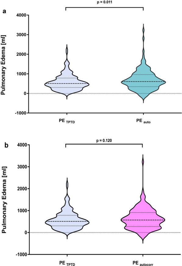
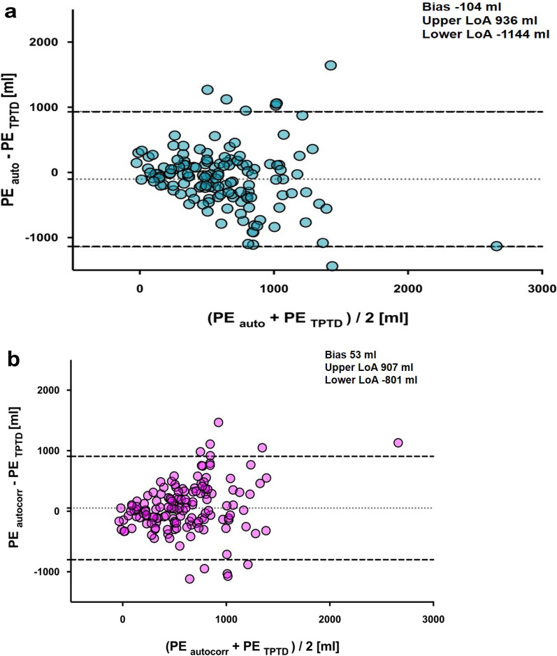
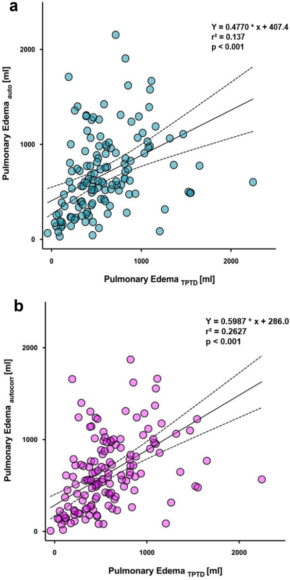
145 comparative measurements of pulmonary edema with TPTD and CT were included in the study. Estimating pulmonary edema using either automated lung segmentation on CT or TPTD showed a low bias overall (- 104 ml) but wide levels of agreement (upper: 936 ml, lower: - 1144 ml). In 13% of the analyzed CT scans, the agreement between the segmentation of the AI algorithm and a dedicated investigator was poor. Manual segmentation and automated segmentation adjusted for contrast agent did not improve the agreement levels.

CONCLUSIONS

Automated lung segmentation on CT can be considered an unbiased but imprecise measurement of pulmonary edema in mechanically ventilated patients with ARDS.

摘要

背景

急性呼吸窘迫综合征(ARDS)患者的肺水肿通过胸部计算机断层扫描(CT)进行定量分析,因其操作复杂且耗时,尚未在常规诊断中得到验证。因此,用于测量血管外肺水(EVLW)的单指标经肺热稀释(TPTD)技术已应用于临床。人工智能(AI)的发展使得在相对较短的时间内,无需事先接受放射学培训的重症监护医生就能自动分割不均匀肺部的CT图像。然而,与TPTD相比,使用CT自动肺分割来验证肺水肿定量分析的数据较少。

方法

一项回顾性研究(2016年1月至2021年12月)分析了入住曼海姆大学医院麻醉与重症医学科重症监护病房的ARDS患者,这些患者同时接受了胸部CT扫描和使用TPTD的血流动力学监测。通过CT上的手动和自动肺分割来估计肺水肿,然后与通过TPTD测定的EVLW计算出的肺水肿进行比较。

结果

该研究纳入了145次TPTD与CT对肺水肿的对比测量。使用CT自动肺分割或TPTD估计肺水肿总体偏差较低(-104 ml),但一致性水平差异较大(上限:936 ml,下限:-1144 ml)。在13%的分析CT扫描中,AI算法分割与专业研究人员之间的一致性较差。针对造影剂进行调整的手动分割和自动分割并未提高一致性水平。

结论

对于机械通气的ARDS患者,CT自动肺分割可被视为对肺水肿的一种无偏差但不精确的测量方法。

https://cdn.ncbi.nlm.nih.gov/pmc/blobs/1ce4/11531458/f76ec82aafd8/40635_2024_685_Fig3_HTML.jpg
https://cdn.ncbi.nlm.nih.gov/pmc/blobs/1ce4/11531458/d171370249a5/40635_2024_685_Fig1_HTML.jpg
https://cdn.ncbi.nlm.nih.gov/pmc/blobs/1ce4/11531458/0b8ce756a4c8/40635_2024_685_Fig2_HTML.jpg
https://cdn.ncbi.nlm.nih.gov/pmc/blobs/1ce4/11531458/f76ec82aafd8/40635_2024_685_Fig3_HTML.jpg
https://cdn.ncbi.nlm.nih.gov/pmc/blobs/1ce4/11531458/d171370249a5/40635_2024_685_Fig1_HTML.jpg
https://cdn.ncbi.nlm.nih.gov/pmc/blobs/1ce4/11531458/0b8ce756a4c8/40635_2024_685_Fig2_HTML.jpg
https://cdn.ncbi.nlm.nih.gov/pmc/blobs/1ce4/11531458/f76ec82aafd8/40635_2024_685_Fig3_HTML.jpg

相似文献

1
Quantification of pulmonary edema using automated lung segmentation on computed tomography in mechanically ventilated patients with acute respiratory distress syndrome.在机械通气的急性呼吸窘迫综合征患者中,利用计算机断层扫描上的自动肺分割技术对肺水肿进行定量分析。
Intensive Care Med Exp. 2024 Nov 2;12(1):95. doi: 10.1186/s40635-024-00685-w.
2
Quantitative computed tomography in comparison with transpulmonary thermodilution for the estimation of pulmonary fluid status: a clinical study in critically ill patients.定量计算机断层扫描与经肺热稀释法用于评估肺内液体状态的比较:一项重症患者的临床研究
J Clin Monit Comput. 2019 Feb;33(1):5-12. doi: 10.1007/s10877-018-0144-1. Epub 2018 Apr 21.
3
[ASSESSMENT OF EXTRAVASCULAR LUNG WATER BY QUANTITATIVE COMPUTER IMAGE ANALYSIS IN PATIENTS WITH ACUTE RESPIRATORY DISTRESS SYNDROME].[急性呼吸窘迫综合征患者血管外肺水的定量计算机图像分析评估]
Anesteziol Reanimatol. 2015 Mar-Apr;60(2):7-12.
4
B-Lines Scores Derived From Lung Ultrasound Provide Accurate Prediction of Extravascular Lung Water Index: An Observational Study in Critically Ill Patients.肺部超声 B 线评分能准确预测血管外肺水指数:危重症患者的观察性研究。
J Intensive Care Med. 2022 Jan;37(1):21-31. doi: 10.1177/0885066620967655. Epub 2020 Nov 5.
5
Assessing pulmonary permeability by transpulmonary thermodilution allows differentiation of hydrostatic pulmonary edema from ALI/ARDS.通过经肺热稀释法评估肺通透性可区分静水压性肺水肿与急性肺损伤/急性呼吸窘迫综合征。
Intensive Care Med. 2007 Mar;33(3):448-53. doi: 10.1007/s00134-006-0498-6. Epub 2007 Jan 13.
6
Resolution of pulmonary edema with variable mechanical ventilation in a porcine model of acute lung injury.肺水肿在急性肺损伤猪模型中通过可变机械通气得到解决。
Can J Anaesth. 2011 Aug;58(8):740-50. doi: 10.1007/s12630-011-9517-3. Epub 2011 Jun 4.
7
Diagnostic value of transpulmonary thermodilution measurements for acute respiratory distress syndrome in a pig model of septic shock.经肺热稀释测量对感染性休克猪模型急性呼吸窘迫综合征的诊断价值。
J Transl Med. 2022 Dec 23;20(1):617. doi: 10.1186/s12967-022-03793-x.
8
Extravascular lung water measurements in acute respiratory distress syndrome: why, how, and when?急性呼吸窘迫综合征的血管外肺水测量:为何、如何以及何时?
Curr Opin Crit Care. 2018 Jun;24(3):209-215. doi: 10.1097/MCC.0000000000000503.
9
Computed tomography to estimate cardiac preload and extravascular lung water. A retrospective analysis in critically ill patients.使用计算机断层扫描估计心脏前负荷和血管外肺水。危重症患者的回顾性分析。
Scand J Trauma Resusc Emerg Med. 2011 May 23;19:31. doi: 10.1186/1757-7241-19-31.
10
Accurate characterization of extravascular lung water in acute respiratory distress syndrome.急性呼吸窘迫综合征中血管外肺水的准确特征描述
Crit Care Med. 2008 Jun;36(6):1803-9. doi: 10.1097/CCM.0b013e3181743eeb.

本文引用的文献

1
A New Global Definition of Acute Respiratory Distress Syndrome.急性呼吸窘迫综合征的新全球定义。
Am J Respir Crit Care Med. 2024 Jan 1;209(1):37-47. doi: 10.1164/rccm.202303-0558WS.
2
ESICM guidelines on acute respiratory distress syndrome: definition, phenotyping and respiratory support strategies.急性呼吸窘迫综合征 ESICM 指南:定义、表型和呼吸支持策略。
Intensive Care Med. 2023 Jul;49(7):727-759. doi: 10.1007/s00134-023-07050-7. Epub 2023 Jun 16.
3
Effects of changes in veno-venous extracorporeal membrane oxygenation blood flow on the measurement of intrathoracic blood volume and extravascular lung water index: a prospective interventional study.
变化的静脉-静脉体外膜肺氧合血流对胸腔血容量和血管外肺水指数测量的影响:一项前瞻性干预研究。
J Clin Monit Comput. 2023 Apr;37(2):599-607. doi: 10.1007/s10877-022-00931-0. Epub 2022 Oct 25.
4
Semi-quantitative evaluation of chest computed tomography for coronavirus disease 2019 in a critical care unit: A case-control study.重症监护病房 2019 冠状病毒病胸部计算机断层扫描的半定量评估:病例对照研究。
Medicine (Baltimore). 2022 Sep 16;101(37):e30655. doi: 10.1097/MD.0000000000030655.
5
Automated Quantification of Inflamed Lung Regions in Chest CT by UNet++ and SegCaps: A Comparative Analysis in COVID-19 Cases.基于 UNet++ 和 SegCaps 的胸部 CT 肺部炎症区域自动量化:COVID-19 病例的对比分析。
Annu Int Conf IEEE Eng Med Biol Soc. 2022 Jul;2022:3785-3788. doi: 10.1109/EMBC48229.2022.9870901.
6
Different Lung Parenchyma Quantification Using Dissimilar Segmentation Software: A Multi-Center Study for COVID-19 Patients.使用不同分割软件对肺实质进行不同量化:一项针对新冠肺炎患者的多中心研究
Diagnostics (Basel). 2022 Jun 20;12(6):1501. doi: 10.3390/diagnostics12061501.
7
Effects of different positive end-expiratory pressure titration strategies during prone positioning in patients with acute respiratory distress syndrome: a prospective interventional study.急性呼吸窘迫综合征患者俯卧位时不同呼气末正压滴定策略的效果:一项前瞻性干预研究。
Crit Care. 2022 Mar 26;26(1):82. doi: 10.1186/s13054-022-03956-8.
8
Personalized mechanical ventilation in acute respiratory distress syndrome.急性呼吸窘迫综合征的个性化机械通气。
Crit Care. 2021 Jul 16;25(1):250. doi: 10.1186/s13054-021-03686-3.
9
Increased extravascular lung water index (EVLWI) reflects rapid non-cardiogenic oedema and mortality in COVID-19 associated ARDS.血管外肺水指数(EVLWI)增加反映了 COVID-19 相关 ARDS 的快速非心源性水肿和死亡率。
Sci Rep. 2021 Jun 1;11(1):11524. doi: 10.1038/s41598-021-91043-3.
10
Multicenter Assessment of CT Pneumonia Analysis Prototype for Predicting Disease Severity and Patient Outcome.多中心 CT 肺炎分析原型评估:预测疾病严重程度和患者转归。
J Digit Imaging. 2021 Apr;34(2):320-329. doi: 10.1007/s10278-021-00430-9. Epub 2021 Feb 25.