Azizian-Farsani Fatemeh, Weixelbaumer Katrin, Mascher Daniel, Klang Andrea, Högler Sandra, Dinhopl Nora, Bauder Barbara, Weissenböck Herbert, Tichy Alexander, Schmidt Peter, Mascher Hermann, Osuchowski Marcin F
Ludwig Boltzmann Institute for Traumatology, The Research Center in Cooperation with AUVA, Donaueschingenstraße 13, 1200, Vienna, Austria.
pharm-analyt Labor GmbH, Baden, Austria.
Intensive Care Med Exp. 2024 Nov 4;12(1):96. doi: 10.1186/s40635-024-00688-7.
Septic encephalopathy is frequent but its pathophysiology is enigmatic. We studied expression of neurotransmitters, inflammation and integrity of the blood-brain barrier (BBB) in several brain regions during abdominal sepsis. We compared mice with either lethal or surviving phenotype in the first 4 sepsis days. Mature CD-1 females underwent cecal ligation and puncture (CLP). Body temperature (BT) was measured daily and predicted-to-die (within 24 h) mice (for P-DIE; BT < 28 °C) were sacrificed together (1:1 ratio) with mice predicted-to-survive (P-SUR; BT > 35 °C), and healthy controls (CON). Brains were dissected into neocortex, cerebellum, midbrain, medulla, striatum, hypothalamus and hippocampus.
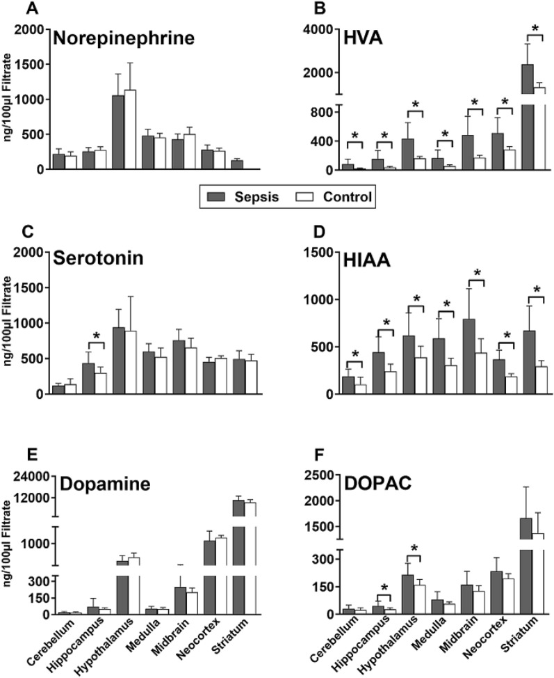
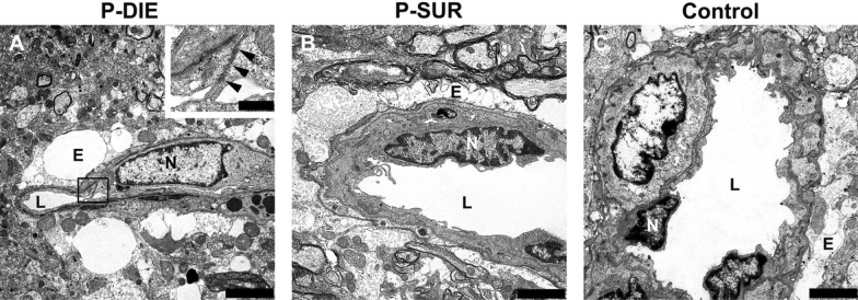
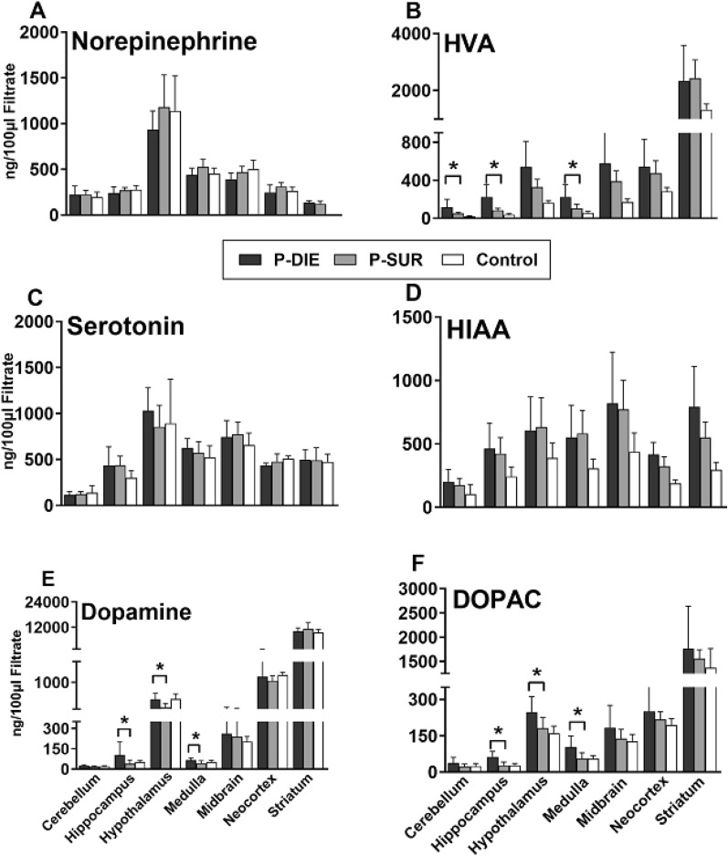
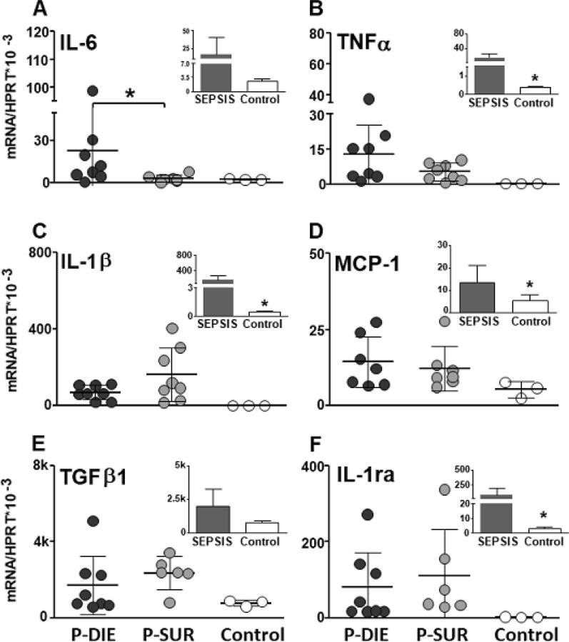
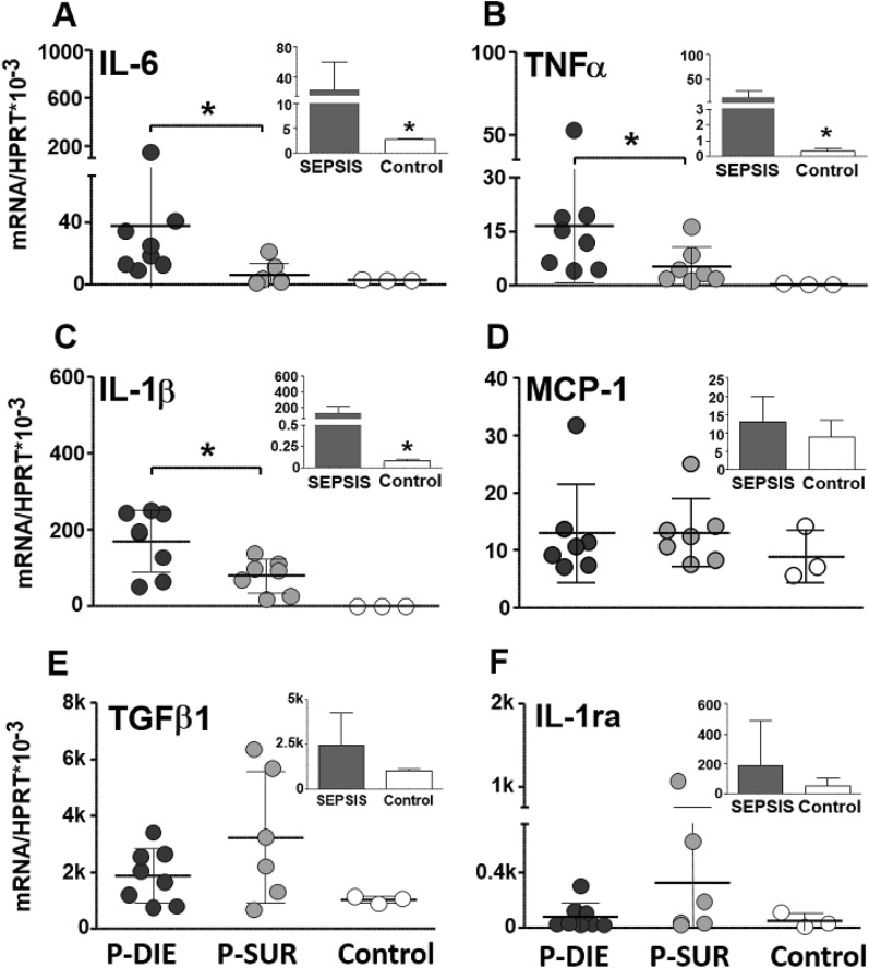
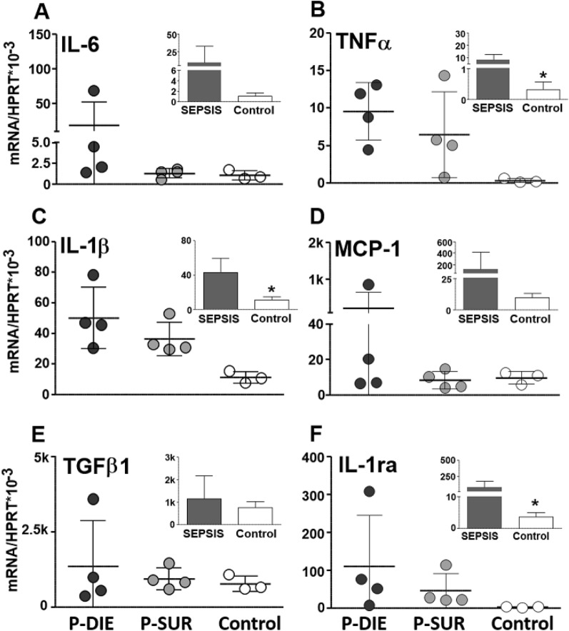
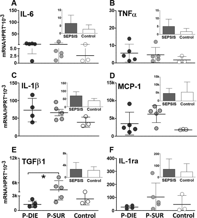
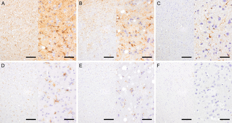
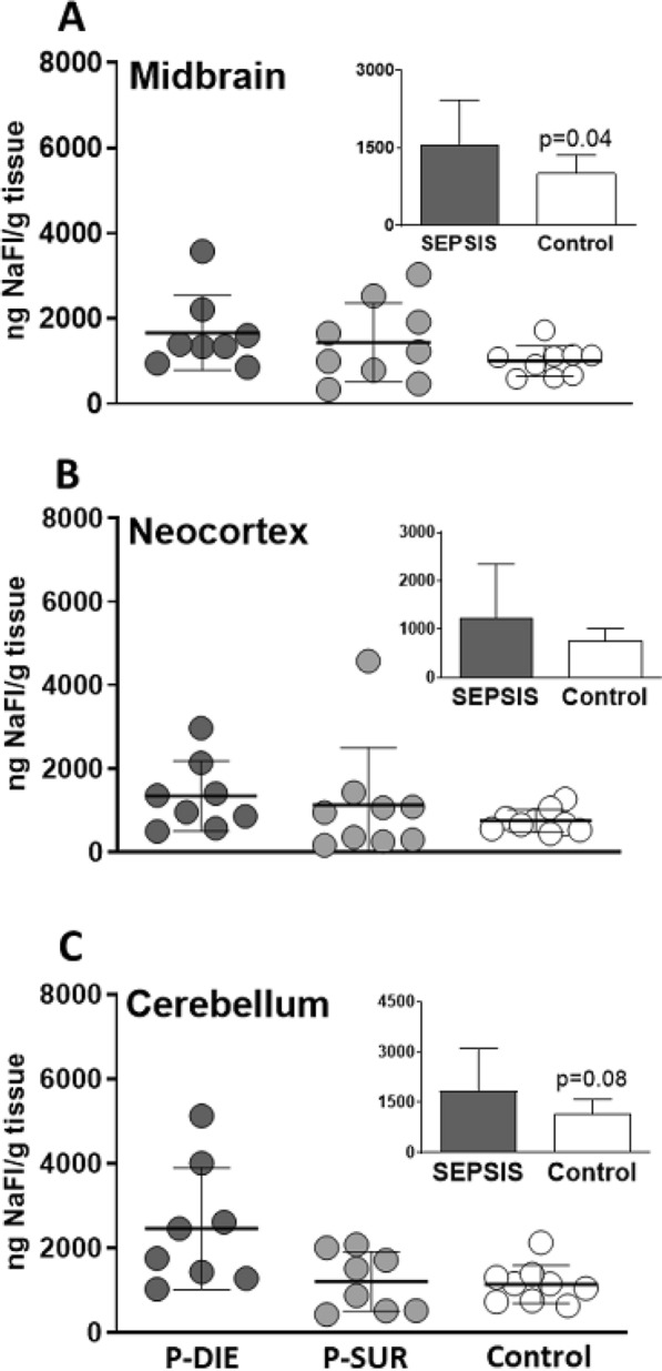
CLP mice showed an up to threefold rise of serotonin in the hippocampus, 5-hydroxyindoleacetic and homovanillic acid (HVA) in nearly all regions vs. CON. Compared to P-SUR, P-DIE mice showed a 1.7 to twofold rise of HVA (386 ng/g of tissue), dopamine (265 ng/g) and 3,4-Dihydroxyphenylacetic acid (DOPAC; 140 ng/g) in the hippocampus, hypothalamus and medulla (174, 156, 82 ng/g of tissue, respectively). CLP increased expression of TNFα, IL-1β and IL-6 mRNA by several folds in the midbrain, cerebellum and hippocampus versus CON. The same cytokines were further elevated in P-DIE vs P-SUR in the midbrain and cerebellum. Activation of astrocytes and microglia was robust across regions but remained typically phenotype independent. There was a similar influx of sodium fluorescein across the BBB in both P-DIE and P-SUR mice.
Compared to survivors, the lethal phenotype induced a stronger deregulation of amine metabolism and cytokine expression in selected brain regions, but the BBB permeability remained similar regardless of the predicted outcome.
脓毒症性脑病很常见,但其病理生理学仍不清楚。我们研究了腹部脓毒症期间几个脑区神经递质的表达、炎症反应及血脑屏障(BBB)的完整性。我们比较了脓毒症最初4天内具有致死或存活表型的小鼠。成年CD-1雌性小鼠接受盲肠结扎和穿刺(CLP)。每天测量体温(BT),将预测死亡(24小时内)的小鼠(P-DIE;BT<28℃)与预测存活的小鼠(P-SUR;BT>35℃)及健康对照(CON)按1:1的比例一同处死。将大脑解剖为新皮质、小脑、中脑、延髓、纹状体、下丘脑和海马体。
与CON相比,CLP小鼠海马体中的5-羟色胺升高了两倍,几乎所有脑区的5-羟吲哚乙酸和高香草酸(HVA)均升高。与P-SUR相比,P-DIE小鼠海马体、下丘脑和延髓中的HVA(386 ng/g组织)、多巴胺(265 ng/g)和3,4-二羟基苯乙酸(DOPAC;140 ng/g)分别升高了1.7至两倍(分别为174、156、82 ng/g组织)。与CON相比,CLP使中脑、小脑和海马体中TNFα、IL-1β和IL-6 mRNA的表达增加了数倍。在中脑和小脑中,与P-SUR相比,P-DIE中相同细胞因子进一步升高。星形胶质细胞和小胶质细胞在所有脑区均被强烈激活,但通常仍与表型无关。P-DIE和P-SUR小鼠血脑屏障对荧光素钠的摄取相似。
与幸存者相比,致死表型在选定脑区引起更强的胺代谢和细胞因子表达失调,但无论预测结果如何,血脑屏障通透性保持相似。