Liverpool Ocular Oncology Research Group, Department of Eye and Vision Science, Institute of Life Course and Medical Science, University of Liverpool, 3rd Floor William Henry Duncan Building, West Derby Street, Liverpool, L7 8TX, UK.
Liverpool Clinical Laboratories, Liverpool University Hospital Foundation Trust, Liverpool, UK.
Sci Rep. 2024 Nov 5;14(1):26811. doi: 10.1038/s41598-024-78171-2.
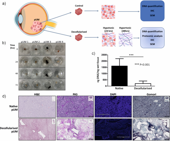
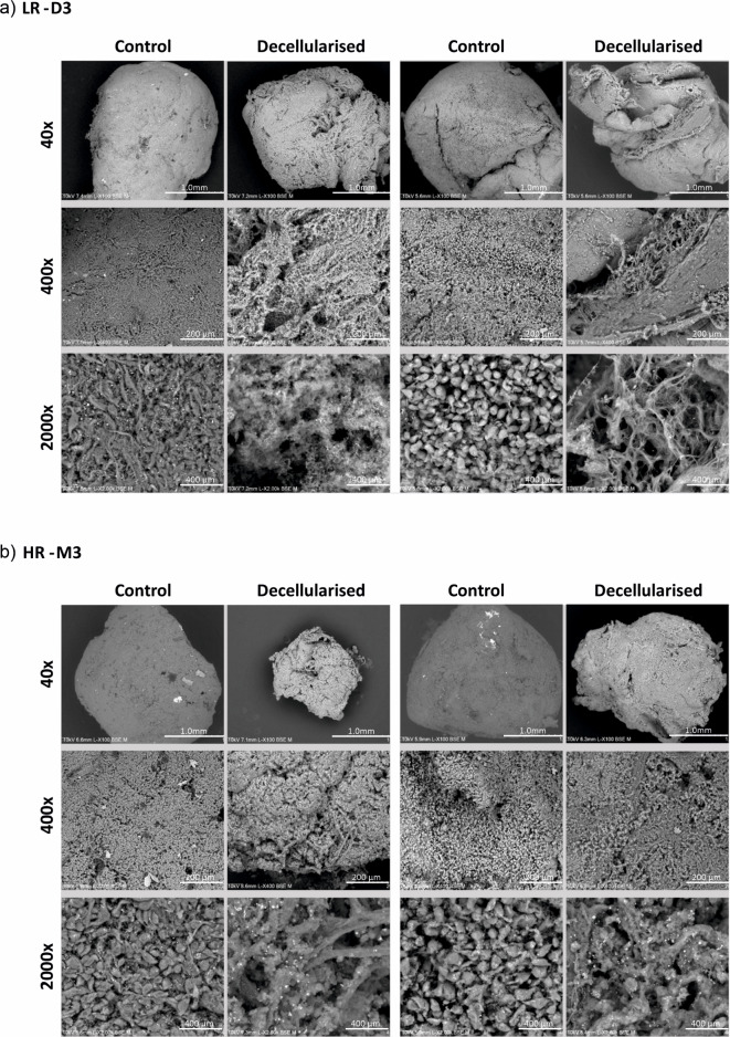
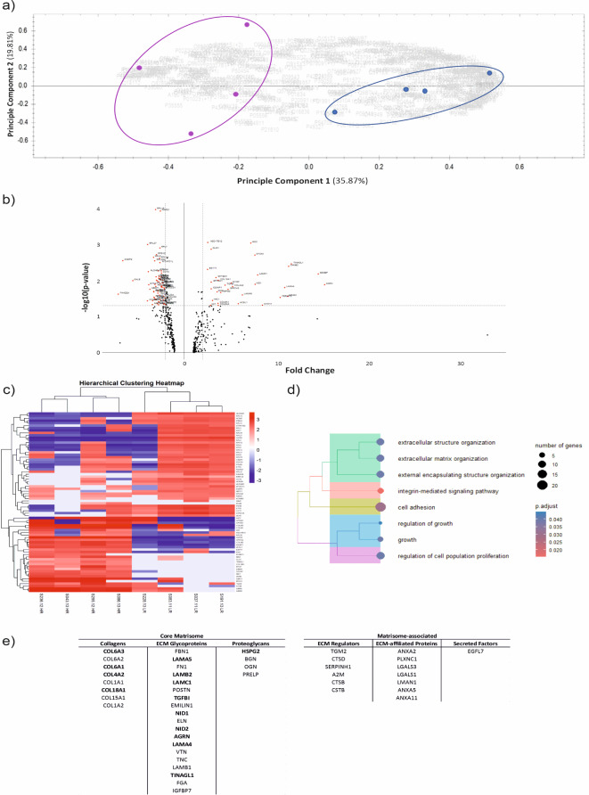
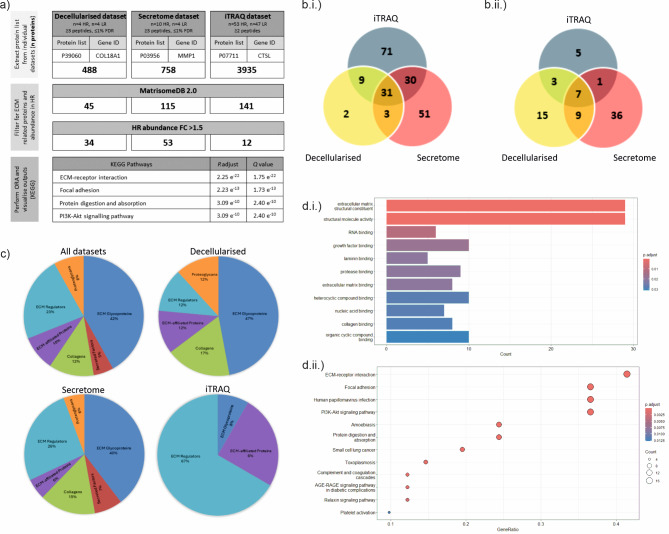
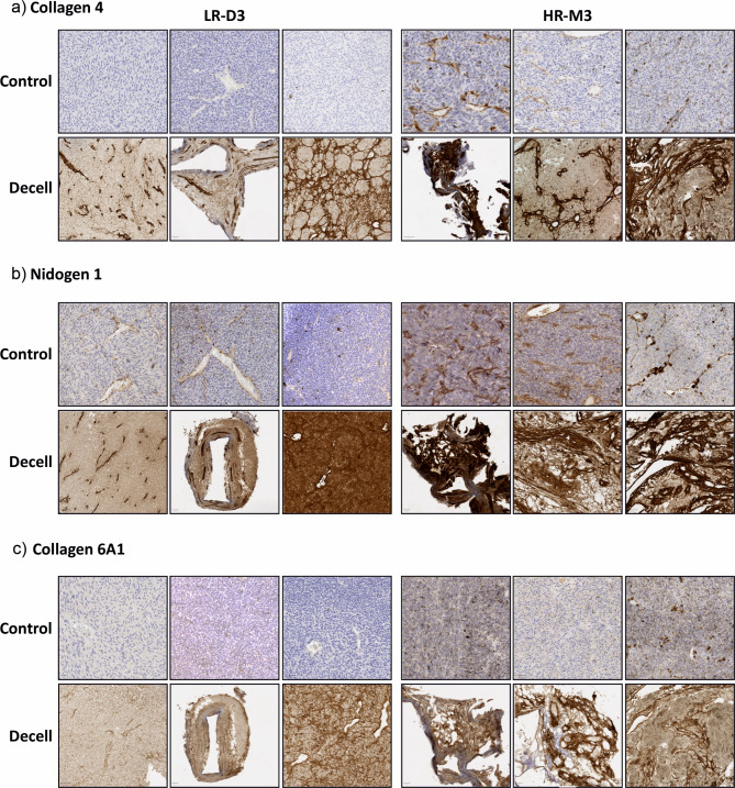
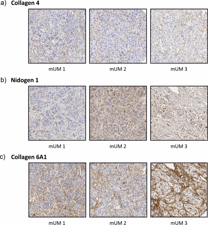
Uveal melanoma (UM) is a rare aggressive intraocular tumour that spreads most commonly to the liver in tumours with loss of one copy of chromosome 3 (HR-M3); current treatments for metastatic disease remain largely ineffective. Pre-clinical research is increasingly using three-dimensional models that better recapitulate the tumour microenvironment (TME). One aspect of the TME is the acellular extracellular matrix (ECM) that influences cell proliferation, migration and response to therapy. Although commercial matrices are used in culture, the composition and biochemical properties may not be representative of the tumour ECM in vivo. This study identifies UM metastatic risk specific ECM proteins by developing methodology for decellularisation of low- and high- metastatic risk tissue samples (LR-D3 vs. HR-M3). Proteomic analysis revealed a matrisome signature of 34 core ECM and ECM-associated proteins upregulated in HR-M3 UM. Combining additional UM secretome and whole cell iTRAQ proteomic datasets revealed enriched GO and KEGG pathways including 'regulating ECM binding' and 'PI3K/Akt signalling'. Structural analyses of decellularised matrices revealed microarchitecture of differing fibre density and expression differences in collagen 4, collagen 6A1 and nidogen 1, between metastatic risk groups. This approach is a powerful tool for the generation of ECM matrices relevant to high metastatic risk UM.
葡萄膜黑色素瘤 (UM) 是一种罕见的侵袭性眼内肿瘤,最常见于失去 3 号染色体 (HR-M3) 拷贝的肿瘤转移到肝脏;目前转移性疾病的治疗方法仍然效果不佳。临床前研究越来越多地使用能够更好地再现肿瘤微环境 (TME) 的三维模型。TME 的一个方面是无细胞细胞外基质 (ECM),它影响细胞增殖、迁移和对治疗的反应。尽管商业基质用于培养,但组成和生化特性可能与体内肿瘤 ECM 不具有代表性。本研究通过开发低转移风险 (LR-D3) 和高转移风险 (HR-M3) 组织样本的去细胞化方法,确定了 UM 转移性风险特异性 ECM 蛋白。蛋白质组学分析显示,HR-M3 UM 中上调了 34 种核心 ECM 和 ECM 相关蛋白的基质组学特征。结合其他 UM 分泌组和全细胞 iTRAQ 蛋白质组数据集,揭示了富含 GO 和 KEGG 途径,包括“调节 ECM 结合”和“PI3K/Akt 信号传导”。去细胞化基质的结构分析揭示了不同纤维密度的微观结构以及胶原 4、胶原 6A1 和巢蛋白 1 之间在转移风险组之间的表达差异。这种方法是生成与高转移风险 UM 相关的 ECM 基质的有力工具。