• 文献检索
  • 文档翻译
  • 深度研究
  • 学术资讯
  • Suppr Zotero 插件Zotero 插件
  • 邀请有礼
  • 套餐&价格
  • 历史记录
应用&插件
Suppr Zotero 插件Zotero 插件浏览器插件Mac 客户端Windows 客户端微信小程序
定价
高级版会员购买积分包购买API积分包
服务
文献检索文档翻译深度研究API 文档MCP 服务
关于我们
关于 Suppr公司介绍联系我们用户协议隐私条款
关注我们

Suppr 超能文献

核心技术专利:CN118964589B侵权必究
粤ICP备2023148730 号-1Suppr @ 2026

文献检索

告别复杂PubMed语法,用中文像聊天一样搜索,搜遍4000万医学文献。AI智能推荐,让科研检索更轻松。

立即免费搜索

文件翻译

保留排版,准确专业,支持PDF/Word/PPT等文件格式,支持 12+语言互译。

免费翻译文档

深度研究

AI帮你快速写综述,25分钟生成高质量综述,智能提取关键信息,辅助科研写作。

立即免费体验

用于共聚焦可视化单个致密核心囊泡的基因编码标记物。

Genetically-encoded markers for confocal visualization of single dense core vesicles.

作者信息

Yu Junwei, Zhang Yunpeng, Clements Kelsey, Chen Nannan, Griffith Leslie C

机构信息

Department of Biology, Volen National Center for Complex Systems, Brandeis University, Waltham, MA 02454-9110, USA.

These authors contributed equally.

出版信息

Res Sq. 2024 Oct 21:rs.3.rs-5021271. doi: 10.21203/rs.3.rs-5021271/v1.

DOI:10.21203/rs.3.rs-5021271/v1
PMID:39502772
原文链接:https://pmc.ncbi.nlm.nih.gov/articles/PMC11537351/
Abstract

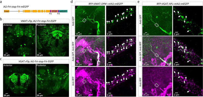
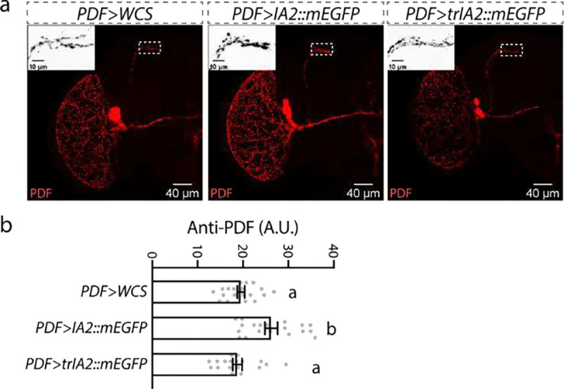
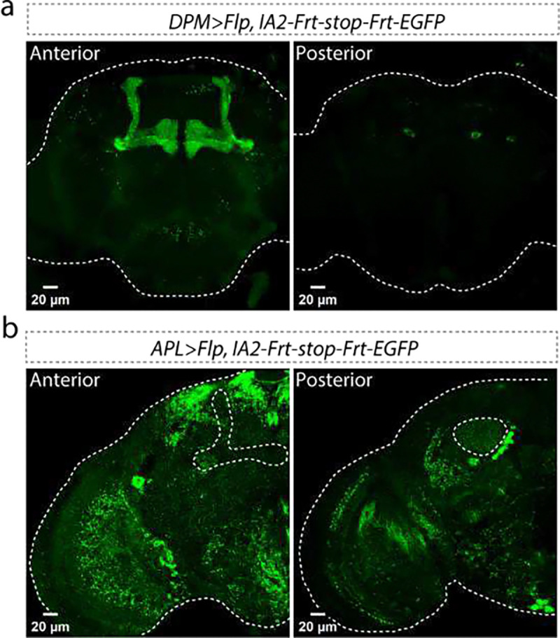
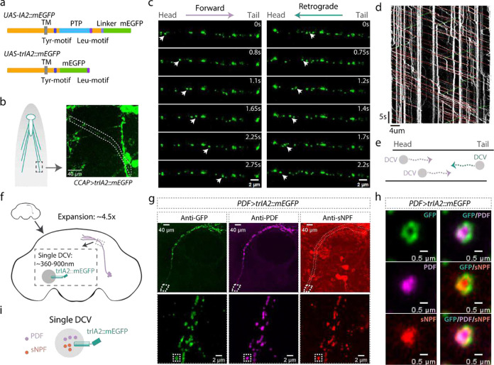
Neuronal dense core vesicles (DCVs) store and release a diverse array of neuromodulators, trophic factors and bioamines. The analysis of single DCVs has largely been possible only using electron microscopy, which makes understanding cargo segregation and DCV heterogeneity difficult. To address these limitations, we developed genetically-encoded markers for DCVs that can be used in combination with standard immunohistochemistry and expansion microscopy, to enable single-vesicle resolution with confocal microscopy.

摘要

神经元致密核心囊泡(DCV)储存并释放多种神经调质、营养因子和生物胺。对单个DCV的分析在很大程度上仅能通过电子显微镜进行,这使得理解货物分选和DCV异质性变得困难。为了解决这些局限性,我们开发了用于DCV的基因编码标记物,其可与标准免疫组织化学和扩展显微镜结合使用,以实现共聚焦显微镜下的单囊泡分辨率。

https://cdn.ncbi.nlm.nih.gov/pmc/blobs/83e0/11537351/ffd066c9c4d0/nihpp-rs5021271v1-f0002.jpg
https://cdn.ncbi.nlm.nih.gov/pmc/blobs/83e0/11537351/87b023b53998/nihpp-rs5021271v1-f0003.jpg
https://cdn.ncbi.nlm.nih.gov/pmc/blobs/83e0/11537351/263440903b9b/nihpp-rs5021271v1-f0004.jpg
https://cdn.ncbi.nlm.nih.gov/pmc/blobs/83e0/11537351/4f4b62a7ec25/nihpp-rs5021271v1-f0005.jpg
https://cdn.ncbi.nlm.nih.gov/pmc/blobs/83e0/11537351/fb73867c3879/nihpp-rs5021271v1-f0006.jpg
https://cdn.ncbi.nlm.nih.gov/pmc/blobs/83e0/11537351/b89d558d05ca/nihpp-rs5021271v1-f0007.jpg
https://cdn.ncbi.nlm.nih.gov/pmc/blobs/83e0/11537351/b3e242594843/nihpp-rs5021271v1-f0008.jpg
https://cdn.ncbi.nlm.nih.gov/pmc/blobs/83e0/11537351/e6af022dfb3e/nihpp-rs5021271v1-f0009.jpg
https://cdn.ncbi.nlm.nih.gov/pmc/blobs/83e0/11537351/bcc9fd69c867/nihpp-rs5021271v1-f0010.jpg
https://cdn.ncbi.nlm.nih.gov/pmc/blobs/83e0/11537351/e41d29eb40af/nihpp-rs5021271v1-f0001.jpg
https://cdn.ncbi.nlm.nih.gov/pmc/blobs/83e0/11537351/ffd066c9c4d0/nihpp-rs5021271v1-f0002.jpg

相似文献

1
Genetically-encoded markers for confocal visualization of single dense core vesicles.用于共聚焦可视化单个致密核心囊泡的基因编码标记物。
Res Sq. 2024 Oct 21:rs.3.rs-5021271. doi: 10.21203/rs.3.rs-5021271/v1.
2
Genetically-encoded markers for confocal visualization of single dense core vesicles.用于共聚焦可视化单个致密核心囊泡的基因编码标记物。
bioRxiv. 2024 Oct 8:2024.10.07.617131. doi: 10.1101/2024.10.07.617131.
3
Genetically-encoded markers for confocal visualization of single dense core vesicles.用于共聚焦可视化单个致密核心囊泡的基因编码标记物。
Commun Biol. 2025 Mar 7;8(1):383. doi: 10.1038/s42003-025-07829-y.
4
Synapsin Is Required for Dense Core Vesicle Capture and cAMP-Dependent Neuropeptide Release.突触结合蛋白对于致密核心囊泡的捕获和 cAMP 依赖性神经肽释放是必需的。
J Neurosci. 2021 May 12;41(19):4187-4201. doi: 10.1523/JNEUROSCI.2631-20.2021. Epub 2021 Apr 5.
5
How old are dense-core vesicles residing in boutons: simulation of the mean age of dense-core vesicles in axonal arbours accounting for resident and transiting vesicle populations.位于终扣中的致密核心囊泡的年龄有多大:对轴突分支中致密核心囊泡平均年龄的模拟,其中考虑了驻留和转运的囊泡群体。
Proc Math Phys Eng Sci. 2020 Sep;476(2241):20200454. doi: 10.1098/rspa.2020.0454. Epub 2020 Sep 30.
6
Maximal Fusion Capacity and Efficient Replenishment of the Dense Core Vesicle Pool in Hippocampal Neurons.海马神经元中最大融合容量和致密核心囊泡池的有效补充。
J Neurosci. 2023 Nov 8;43(45):7616-7625. doi: 10.1523/JNEUROSCI.2251-22.2023. Epub 2023 Oct 18.
7
Deletion of β-Neurexins in Mice Alters the Distribution of Dense-Core Vesicles in Presynapses of Hippocampal and Cerebellar Neurons.小鼠中β-神经连接蛋白的缺失改变了海马体和小脑神经元突触前致密核心囊泡的分布。
Front Neuroanat. 2022 Jan 31;15:757017. doi: 10.3389/fnana.2021.757017. eCollection 2021.
8
The Phosphoprotein Synapsin Ia Regulates the Kinetics of Dense-Core Vesicle Release.磷酸化突触结合蛋白 Ia 调节致密核心囊泡释放的动力学。
J Neurosci. 2021 Mar 31;41(13):2828-2841. doi: 10.1523/JNEUROSCI.2593-19.2021. Epub 2021 Feb 25.
9
Pool size estimations for dense-core vesicles in mammalian CNS neurons.哺乳动物中枢神经系统神经元中致密核心囊泡的池大小估计。
EMBO J. 2018 Oct 15;37(20). doi: 10.15252/embj.201899672. Epub 2018 Sep 5.
10
Simulation of a sudden drop-off in distal dense core vesicle concentration in Drosophila type II motoneuron terminals.果蝇 II 型运动神经元末梢中远心致密核心囊泡浓度的突然下降模拟。
Int J Numer Method Biomed Eng. 2021 Dec;37(12):e3523. doi: 10.1002/cnm.3523. Epub 2021 Sep 8.

本文引用的文献

1
Widespread posttranscriptional regulation of cotransmission.共传递的广泛转录后调控。
Sci Adv. 2023 Jun 2;9(22):eadg9836. doi: 10.1126/sciadv.adg9836.
2
Functional neuroanatomy of monoaminergic systems in the basolateral nuclear complex of the amygdala: Neuronal targets, receptors, and circuits.杏仁体基底外侧核复合体中单胺能系统的功能神经解剖学:神经元靶点、受体和回路。
J Neurosci Res. 2023 Sep;101(9):1409-1432. doi: 10.1002/jnr.25201. Epub 2023 May 11.
3
The emergence and influence of internal states.内部状态的出现和影响。
Neuron. 2022 Aug 17;110(16):2545-2570. doi: 10.1016/j.neuron.2022.04.030. Epub 2022 May 27.
4
Co-packaging of opposing neurotransmitters in individual synaptic vesicles in the central nervous system.中枢神经系统中单个突触小泡中对向神经递质的共包装。
Neuron. 2022 Apr 20;110(8):1371-1384.e7. doi: 10.1016/j.neuron.2022.01.007. Epub 2022 Feb 3.
5
Live Imaging of Axonal Transport in the Motor Neurons of Larvae.幼虫运动神经元轴突运输的实时成像
Bio Protoc. 2017 Dec 5;7(23):e2631. doi: 10.21769/BioProtoc.2631.
6
Extrasynaptic δ-subunit containing GABA receptors.含 δ 亚基的突触外 GABA 受体。
J Integr Neurosci. 2021 Mar 30;20(1):173-184. doi: 10.31083/j.jin.2021.01.284.
7
Inhibitory Interactions and Columnar Inputs to an Object Motion Detector in Drosophila.果蝇中物体运动检测器的抑制性相互作用和柱状输入。
Cell Rep. 2020 Feb 18;30(7):2115-2124.e5. doi: 10.1016/j.celrep.2020.01.061.
8
Expansion microscopy: development and neuroscience applications.扩展显微镜:发展与神经科学应用。
Curr Opin Neurobiol. 2018 Jun;50:56-63. doi: 10.1016/j.conb.2017.12.012. Epub 2018 Jan 6.
9
GABA Signaling and Neuroactive Steroids in Adrenal Medullary Chromaffin Cells.肾上腺髓质嗜铬细胞中的γ-氨基丁酸信号传导与神经活性类固醇
Front Cell Neurosci. 2016 Apr 18;10:100. doi: 10.3389/fncel.2016.00100. eCollection 2016.
10
Optical imaging. Expansion microscopy.光学成像。扩展显微镜技术。
Science. 2015 Jan 30;347(6221):543-8. doi: 10.1126/science.1260088. Epub 2015 Jan 15.