MIT Media Lab, Massachusetts Institute of Technology, Cambridge, MA, USA.
MIT Media Lab, Massachusetts Institute of Technology, Cambridge, MA, USA; Department of Biological Engineering, Massachusetts Institute of Technology, Cambridge, MA, USA; McGovern Institute for Brain Research, Massachusetts Institute of Technology, Cambridge, MA, USA; Department of Brain and Cognitive Sciences, Massachusetts Institute of Technology, Cambridge, MA, USA.
Curr Opin Neurobiol. 2018 Jun;50:56-63. doi: 10.1016/j.conb.2017.12.012. Epub 2018 Jan 6.
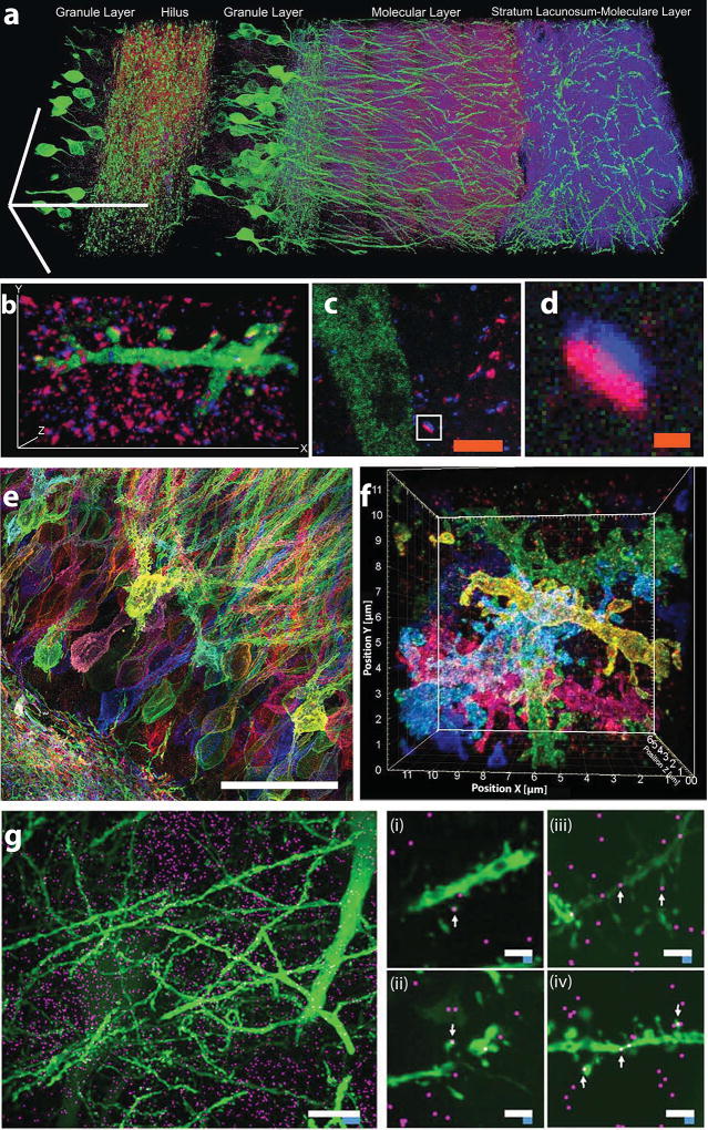
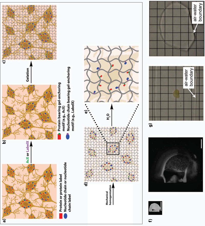
Many neuroscience questions center around understanding how the molecules and wiring in neural circuits mechanistically yield behavioral functions, or go awry in disease states. However, mapping the molecules and wiring of neurons across the large scales of neural circuits has posed a great challenge. We recently developed expansion microscopy (ExM), a process in which we physically magnify biological specimens such as brain circuits. We synthesize throughout preserved brain specimens a dense, even mesh of a swellable polymer such as sodium polyacrylate, anchoring key biomolecules such as proteins and nucleic acids to the polymer. After mechanical homogenization of the specimen-polymer composite, we add water, and the polymer swells, pulling biomolecules apart. Due to the larger separation between molecules, ordinary microscopes can then perform nanoscale resolution imaging. We here review the ExM technology as well as applications to the mapping of synapses, cells, and circuits, including deployment in species such as Drosophila, mouse, non-human primate, and human.
许多神经科学问题都集中在理解神经回路中的分子和连接如何在机械上产生行为功能,或者在疾病状态下出现异常。然而,在神经回路的大尺度上绘制神经元的分子和连接已经构成了巨大的挑战。我们最近开发了扩展显微镜(ExM)技术,这是一种物理放大生物样本(如脑回路)的过程。我们在保存完好的脑样本中合成了一种可膨胀的聚合物(如聚丙烯酸钠)的密集、均匀的网格,将关键的生物分子(如蛋白质和核酸)锚定在聚合物上。在标本-聚合物复合材料进行机械匀浆后,我们加入水,聚合物膨胀,将生物分子拉开。由于分子之间的间隔更大,普通显微镜可以进行纳米级分辨率的成像。在这里,我们回顾了 ExM 技术以及它在突触、细胞和回路测绘中的应用,包括在果蝇、老鼠、非人类灵长类动物和人类等物种中的部署。