Rohowetz Landon J, Staropoli Patrick, da Cruz Natasha F S, Mendoza Carlos, Starke Robert M, Morcos Jacques J, Berrocal Audina M
Department of Ophthalmology, Bascom Palmer Eye Institute, 900 NW 17th St., Miami, FL, USA.
Department of Neurological Surgery, University of Miami, Jackson Health System, 1095 Northwest 14th Ter., Miami, FL, 33136, USA.
Am J Ophthalmol Case Rep. 2024 Oct 31;36:102214. doi: 10.1016/j.ajoc.2024.102214. eCollection 2024 Dec.
To describe the clinical findings in an 11-year-old male with a history of hemifacial microsomia presenting with ocular ischemic syndrome secondary to large cerebral aneurysms.
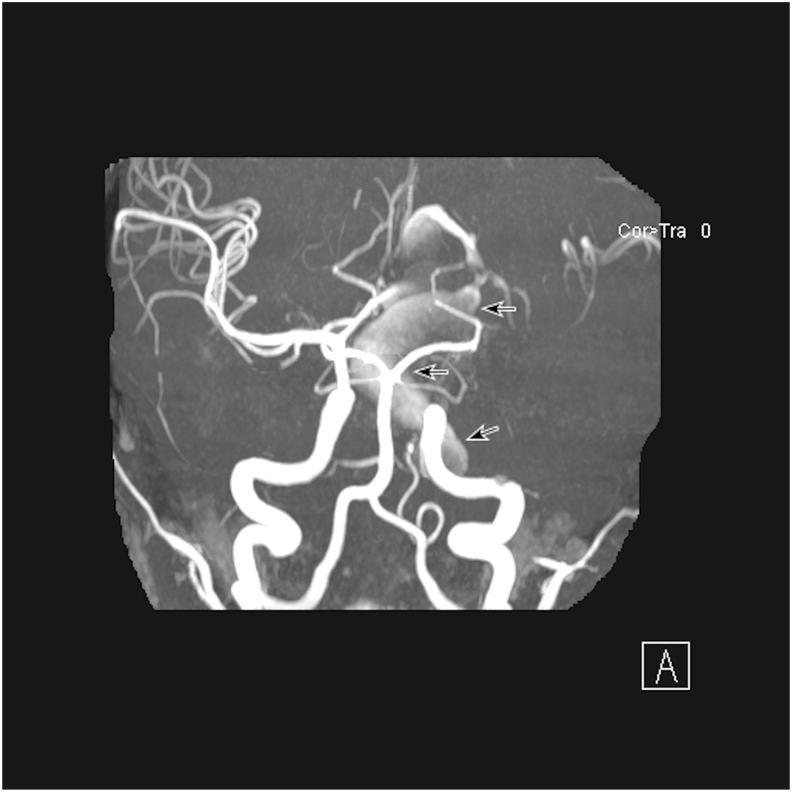
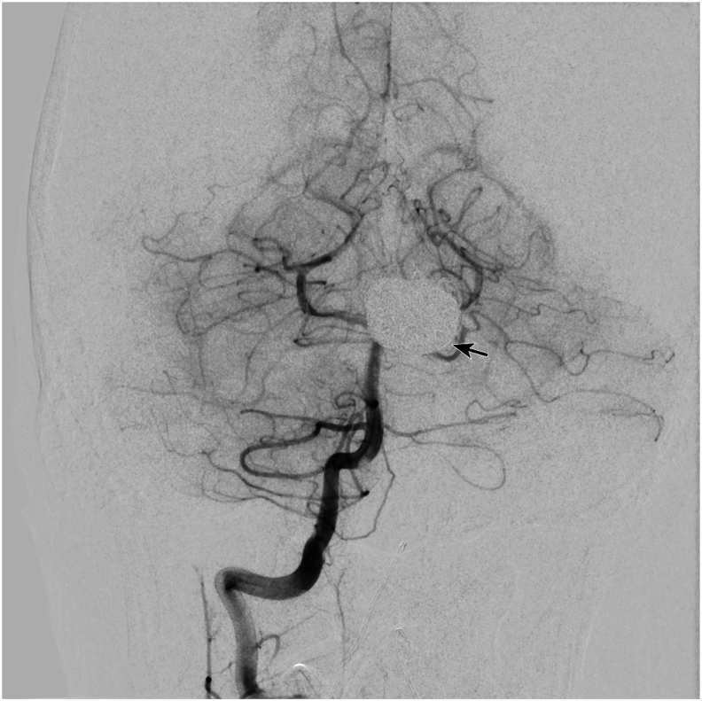
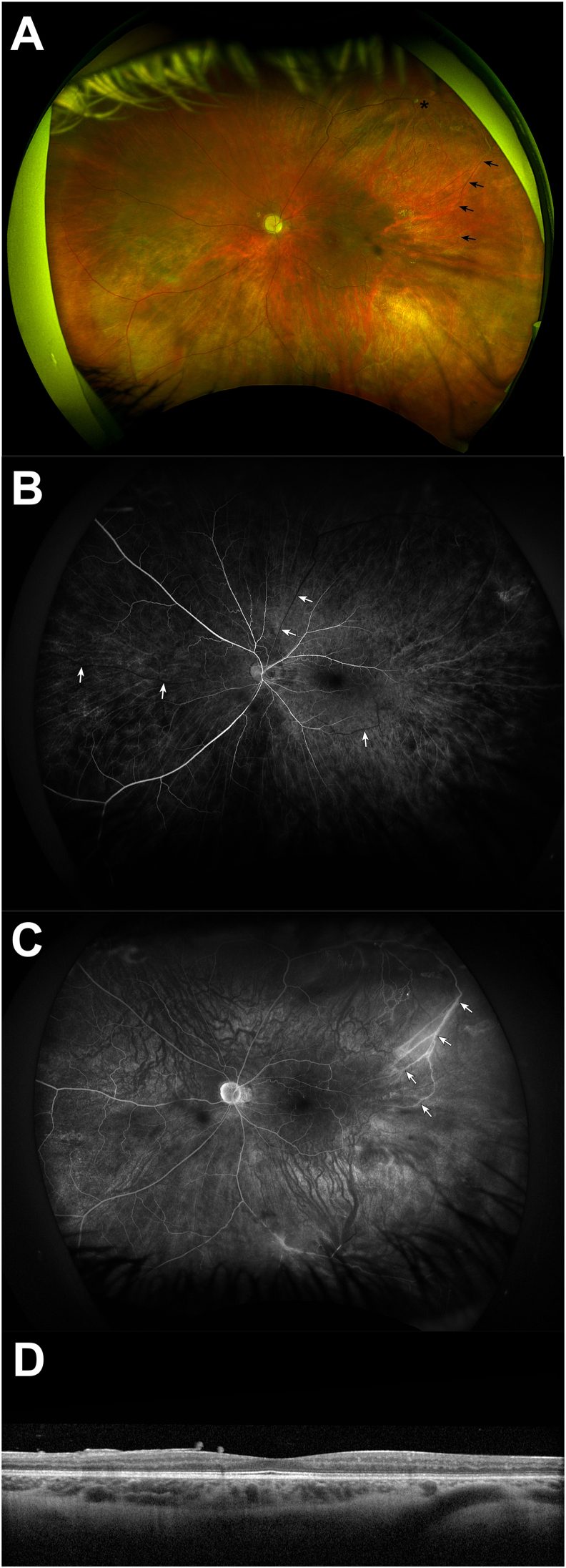
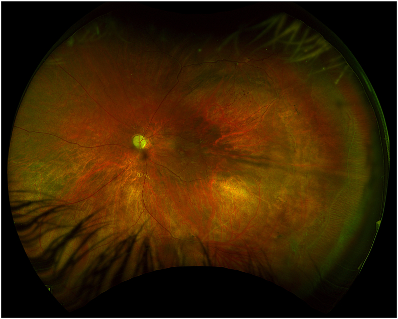
An 11-year-old male with a history of hemifacial microsomia presented to the Bascom Palmer Eye Institute Emergency Department complaining of nausea, diarrhea, headache, and decreased vision in the left eye. Visual acuity was light perception in the left eye and intraocular pressure was within normal limits. Gonioscopy revealed the presence of diffuse neovascularization of the angle. Posterior segment examination revealed mild vitreous hemorrhage, optic disc pallor, preretinal hemorrhage, generalized arteriolar narrowing, retinal microaneurysms, and abnormal arteriovenous communications with branching retinal vessels. Fluorescein angiography demonstrated patchy and delayed choroidal filling, a prolonged venous filling time, arteriolar attenuation, and vascular staining consistent with ocular ischemic syndrome. Magnetic resonance angiography was obtained which revealed large left internal carotid and anterior cerebral artery aneurysms. The patient underwent successful cerebral revascularization via bypass, ligation, clipping, and coiling procedures. At postoperative year 1, there was no evidence of ocular neovascularization and visual acuity remained light perception.
Ocular ischemic syndrome is uncommon in children but may occur with any cause of ocular hypoperfusion. Hemifacial microsomia is a rare congenital disorder of craniofacial development caused by a vascular event in utero affecting the first and second branchial arches. This case demonstrates a rare cause of ocular ischemic syndrome and illustrates the potential for the development of clinically significant vascular abnormalities in patients with disorders of craniofacial development.
描述一名11岁男性的临床症状,该男性有半侧颜面短小畸形病史,现因大脑大动脉瘤继发眼部缺血综合征就诊。
一名有半侧颜面短小畸形病史的11岁男性到巴斯科姆·帕尔默眼科研究所急诊科就诊,主诉恶心、腹泻、头痛及左眼视力下降。左眼视力为光感,眼压在正常范围内。前房角镜检查发现房角有弥漫性新生血管形成。眼后段检查发现轻度玻璃体出血、视盘苍白、视网膜前出血、全身性小动脉狭窄、视网膜微动脉瘤以及视网膜血管分支处异常的动静脉交通。荧光素血管造影显示脉络膜充盈斑片状且延迟、静脉充盈时间延长、小动脉变细以及与眼部缺血综合征相符的血管染色。磁共振血管造影显示左侧颈内动脉和大脑前动脉有大动脉瘤。患者通过搭桥、结扎、夹闭和栓塞手术成功实现了脑部血管重建。术后第1年,未发现眼部新生血管形成的迹象,视力仍为光感。
眼部缺血综合征在儿童中并不常见,但可由任何导致眼部灌注不足的原因引起。半侧颜面短小畸形是一种罕见的颅面发育先天性疾病,由子宫内影响第一和第二鳃弓的血管事件引起。本病例展示了眼部缺血综合征的一种罕见病因,并说明了颅面发育障碍患者发生具有临床意义的血管异常的可能性。