Waheed-Ullah Qazi, Wilsdon Anna, Abbad Aseel, Rochette Sophie, Bu'Lock Frances, Saed Asma Ali, Hitz Marc-Phillip, Brook J David, Loughna Siobhan
School of Life Sciences, Faculty of Medicine and Health Sciences, University of Nottingham, Nottingham, UK.
East Midlands Congenital Heart Centre, University Hospitals of Leicester NHS Trust, Leicester, UK.
J Anat. 2025 Apr;246(4):616-630. doi: 10.1111/joa.14175. Epub 2024 Nov 18.
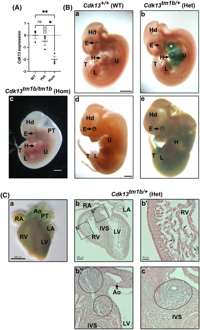
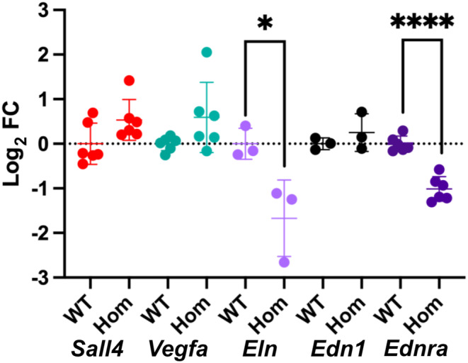
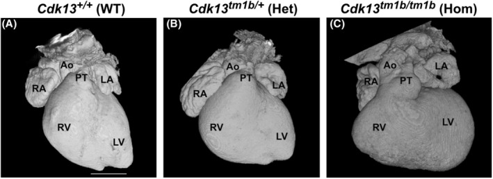
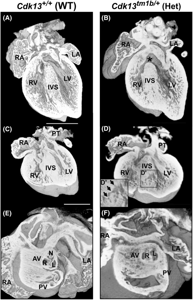
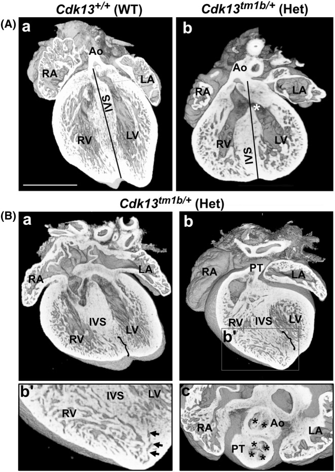
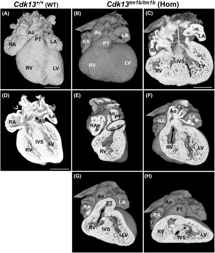
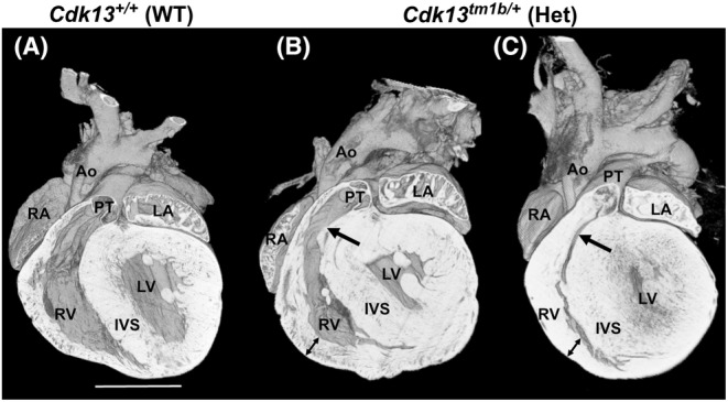
Congenital heart disease (CHD) has an incidence of approximately 1%. Over the last decade, sequencing studies including large cohorts of individuals with CHD have begun to unravel the genetic mechanisms underpinning CHD. This includes the identification of variants in cyclin-dependent kinase 13 (CDK13), in individuals with syndromic CHD. CDK13 encodes a serine/threonine protein kinase. The cyclin partner of CDK13 is cyclin K; this complex is thought to be important in transcription and RNA processing. Pathogenic variants in CDK13 cause CDK13-related disorder in humans, characterised by intellectual disability and developmental delay, recognisable facial features, feeding difficulties and structural brain defects, with 35% of individuals having CHD. To obtain a greater understanding for the role that this essential protein kinase plays in embryonic heart development, we have analysed a presumed loss of function Cdk13 transgenic mouse model (Cdk13). The homozygous mutants were embryonically lethal in most cases by E15.5. X-gal staining showed Cdk13 expression localised to developing facial regions, heart and surrounding areas at E10.5, whereas at E12.5, it was more widely present. In the E15.5 heart, staining was seen throughout. RT-qPCR showed significant reduction in Cdk13 transcript expression in homozygous compared with WT and heterozygous hearts at E10.5 and E12.5. Detailed morphological 3D analysis of embryonic and postnatal hearts was performed using high-resolution episcopic microscopy, which affords a more detailed analysis of structures such as cardiac valve leaflets and endocardial cushions, compared with more traditional histological techniques. We show that both the homozygous and heterozygous Cdk13 mutants exhibit a range of CHD, including ventricular septal defects, bicuspid aortic valve, double outlet right ventricle and atrioventricular septal defects. 100% (n = 4) of homozygous hearts displayed CHD. Differential expression was seen in Cdk13 homozygous mutants for two genes known to be necessary for normal heart development. The types of defects, and the presence of CHD in heterozygous mice (17.02%, n = 8/47), are consistent with the CDK13-related disorder phenotype in humans. This study provides important insights into the effects of reduced function of CDK13 in the mouse heart and contributes to our understanding of the mechanism behind this disorder as a cause of CHD.
先天性心脏病(CHD)的发病率约为1%。在过去十年中,包括大量先天性心脏病患者队列的测序研究已开始揭示先天性心脏病背后的遗传机制。这包括在患有综合征性先天性心脏病的个体中鉴定细胞周期蛋白依赖性激酶13(CDK13)的变异。CDK13编码一种丝氨酸/苏氨酸蛋白激酶。CDK13的细胞周期蛋白伴侣是细胞周期蛋白K;这种复合物被认为在转录和RNA加工中很重要。CDK13中的致病变异会导致人类出现与CDK13相关的疾病,其特征为智力残疾和发育迟缓、可识别的面部特征、喂养困难和结构性脑缺陷,35%的个体患有先天性心脏病。为了更深入了解这种重要的蛋白激酶在胚胎心脏发育中的作用,我们分析了一种推测功能缺失的Cdk13转基因小鼠模型(Cdk13)。在大多数情况下,纯合突变体在胚胎期E15.5时致死。X - gal染色显示,在E10.5时,Cdk13表达定位于发育中的面部区域、心脏及周围区域,而在E12.5时,其分布更为广泛。在E15.5的心脏中,各处均可见染色。RT - qPCR显示,与野生型和杂合子心脏相比,在E10.5和E12.5时,纯合子心脏中Cdk13转录本表达显著降低。使用高分辨率体表显微镜对胚胎期和出生后心脏进行了详细的形态学三维分析,与更传统的组织学技术相比,该技术能对诸如心脏瓣膜小叶和心内膜垫等结构进行更详细的分析。我们发现,纯合子和杂合子Cdk13突变体均表现出一系列先天性心脏病,包括室间隔缺损、二叶式主动脉瓣、右心室双出口和房室间隔缺损。100%(n = 4)的纯合子心脏表现出先天性心脏病。在已知对正常心脏发育必需的两个基因中,Cdk13纯合突变体存在差异表达。杂合子小鼠中缺陷的类型以及先天性心脏病的存在情况(17.02%,n = 8/47)与人类与CDK13相关的疾病表型一致。这项研究为CDK13功能降低对小鼠心脏的影响提供了重要见解,并有助于我们理解这种疾病作为先天性心脏病病因背后的机制。