Department of Diagnostic and Interventional Radiology, University Hospital Zürich, Zürich, Switzerland.
b-rayZ AG, Schlieren, Switzerland.
Eur Radiol Exp. 2024 Nov 18;8(1):129. doi: 10.1186/s41747-024-00527-0.
The presence of a blurred area, depending on its localization, in a mammogram can limit diagnostic accuracy. The goal of this study was to develop a model for automatic detection of blur in diagnostically relevant locations in digital mammography.
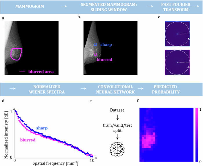
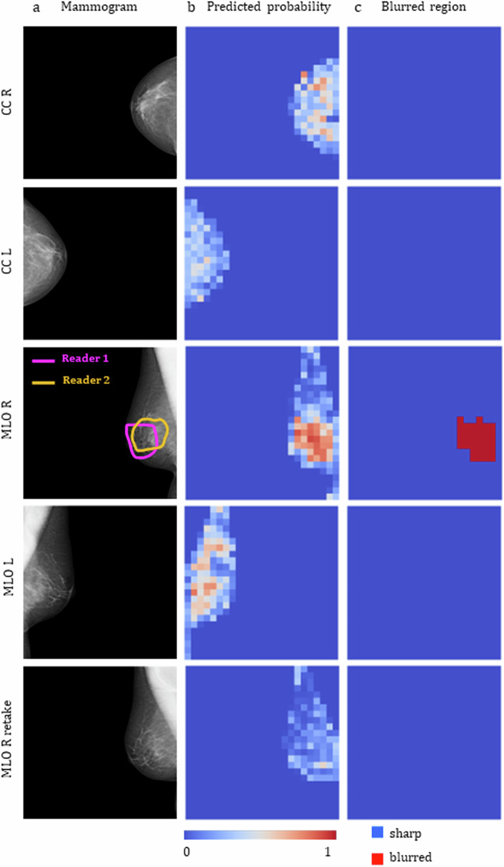
A retrospective dataset consisting of 152 examinations acquired with mammography machines from three different vendors was utilized. The blurred areas were contoured by expert breast radiologists. Normalized Wiener spectra (nWS) were extracted in a sliding window manner from each mammogram. These spectra served as input for a convolutional neural network (CNN) generating the probability of the spectra originating from a blurred region. The resulting blur probability mask, upon thresholding, facilitated the classification of a mammogram as either blurred or sharp. Ground truth for the test set was defined by the consensus of two radiologists.
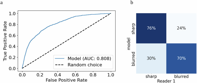
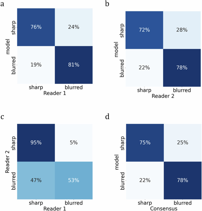
A significant correlation between the view (p < 0.001), as well as between the laterality and the presence of blur (p = 0.004) was identified. The developed model AUROC of 0.808 (95% confidence interval 0.794-0.821) aligned with the consensus in 78% (67-83%) of mammograms classified as blurred. For mammograms classified by consensus as sharp, the model achieved agreement in 75% (67-83%) of them.
A model for blur detection was developed and assessed. The results indicate that a robust approach to blur detection, based on feature extraction in frequency space, tailored to radiologist expertise regarding clinical relevance, could eliminate the subjectivity associated with the visual assessment.
This blur detection model, if implemented in clinical practice, could provide instantaneous feedback to technicians, allowing for prompt mammogram retakes and ensuring that only high-quality mammograms are sent for screening and diagnostic tasks.
Blurring in mammography limits radiologist interpretation and diagnostic accuracy. This objective blur detection tool ensures image quality, and reduces retakes and unnecessary exposures. Wiener spectrum analysis and CNN enabled automated blur detection in mammography.
在乳房 X 光片中,模糊区域的存在(取决于其位置)会限制诊断准确性。本研究的目的是开发一种模型,用于自动检测数字乳房 X 光片中诊断相关位置的模糊。
使用来自三个不同供应商的乳房 X 光机获取的 152 次检查的回顾性数据集。由经验丰富的乳腺放射科医生勾勒出模糊区域。以滑动窗口的方式从每个乳房 X 光片中提取归一化 Wiener 谱(nWS)。这些频谱作为卷积神经网络(CNN)的输入,生成频谱来自模糊区域的概率。经阈值处理后的模糊概率掩模有助于将乳房 X 光片分类为模糊或清晰。测试集的真实情况由两位放射科医生的共识定义。
发现视图之间(p<0.001)以及侧别和模糊之间存在显著相关性(p=0.004)。开发的模型 AUROC 为 0.808(95%置信区间 0.794-0.821),与共识一致,将 78%(67-83%)的乳房 X 光片分类为模糊。对于共识分类为清晰的乳房 X 光片,模型在 75%(67-83%)的乳房 X 光片中达成一致。
开发并评估了一种用于模糊检测的模型。结果表明,基于频率空间中的特征提取,针对放射科医生关于临床相关性的专业知识量身定制的稳健模糊检测方法,可以消除与视觉评估相关的主观性。
如果在临床实践中实施此模糊检测模型,可以为技术人员提供即时反馈,从而可以及时重新拍摄乳房 X 光片,并确保仅发送高质量的乳房 X 光片进行筛查和诊断任务。
乳房 X 光片中的模糊会限制放射科医生的解释和诊断准确性。此客观模糊检测工具可确保图像质量,并减少重拍和不必要的曝光。Wiener 谱分析和 CNN 使乳房 X 光中的自动模糊检测成为可能。