University of Wisconsin-Madison, Department of Medical Physics, Madison, Wisconsin, United States.
CIFICEN (UNCPBA - CICPBA - CONICET), Tandil, Buenos Aires, Argentina.
J Biomed Opt. 2025 Jan;30(Suppl 1):S13709. doi: 10.1117/1.JBO.30.S1.S13709. Epub 2024 Nov 18.
Fluorescence sensing within tissue is an effective tool for tissue characterization; however, the modality and geometry of the image acquisition can alter the observed signal.
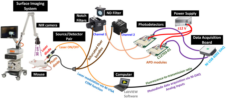
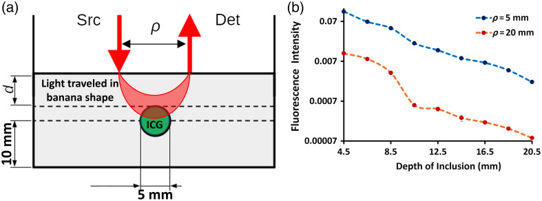
We introduce a novel optical fiber-based system capable of measuring two fluorescent contrast agents through 2 cm of tissue with simple passive electronic switching between the excitation light, simultaneously acquiring fluorescence and excitation data. The goal was to quantify indocyanine green (ICG) and protoporphyrin IX (PpIX) within tissue, and the sampling method was compared with wide-field surface imaging to contrast the value of deep sensing versus surface imaging.
This was achieved by choosing filters for specific wavelengths that were mutually exclusive between ICG and PpIX and coupling these filters to two separate detectors, which allows for direct swapping of the excitation and emission channels by switching the on-time of each excitation laser between 780- and 633-nm wavelengths.
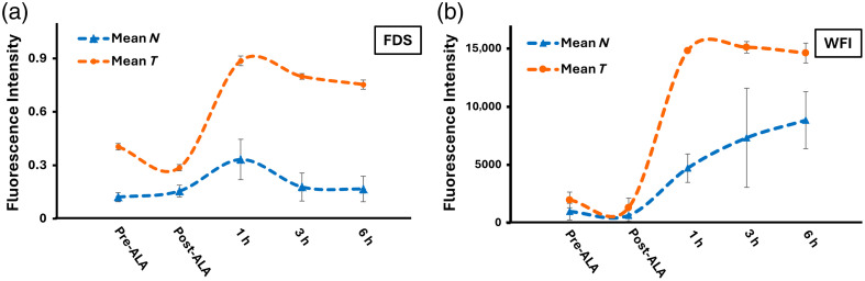
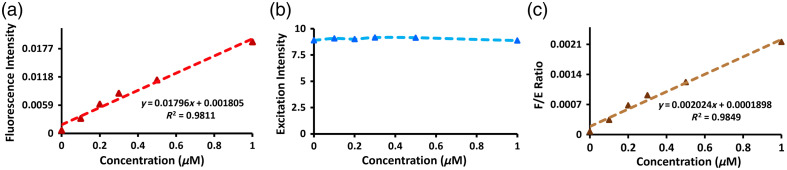
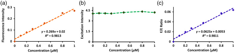
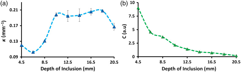
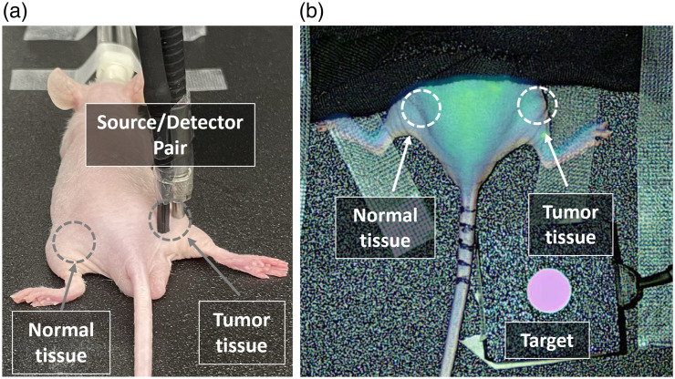
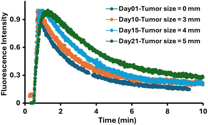
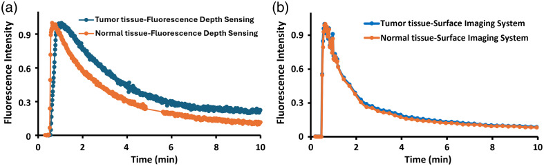
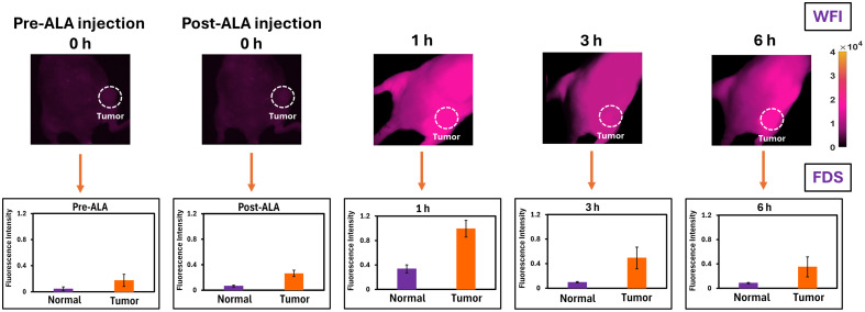
This system was compared with two non-contact surface imaging systems for both ICG and PpIX, which revealed that the fluorescence depth sensing system was superior in its ability to resolve kinetics differences in deeper tissues that would normally be dominated by strong signals from skin and other surface tissues. Specifically, the system was tested using pancreatic adenocarcinoma tumors injected into murine models, which were imaged at several time points throughout tumor growth to its diameter. This demonstrated the system's capability to track longitudinal changes in ICG and PpIX kinetics that result from tumor growth and development, with larger tumors showing sluggish uptake and clearance of ICG, which was not observable with surface imaging. Similarly, PpIX was quantified, which showed slower kinetics over different time points, and was further compared with the wide-filed imager. These results were further validated through depth measurements in tissue phantoms and model-based interpretation.
This fluorescence depth sensing system can be used to sample the interior blood flow characteristics by ICG sensing of tissue as deep as 20 mm into the tissue with sensitivity to kinetics that are superior to surface imaging and may be combined with other imaging modalities such as ultrasound to provide guided deep fluorescence measurements.
组织内的荧光传感是组织特征化的有效工具;然而,模态和图像采集的几何形状会改变观察到的信号。
我们引入了一种新的基于光纤的系统,该系统能够通过简单的被动电子切换在激发光之间测量 2cm 组织内的两种荧光对比剂,同时获取荧光和激发数据。目标是量化组织内的吲哚菁绿(ICG)和原卟啉 IX(PpIX),并将采样方法与宽场表面成像进行比较,以对比深层传感与表面成像的价值。
通过选择特定波长的滤波器来实现这一点,这些滤波器在 ICG 和 PpIX 之间是相互排斥的,并将这些滤波器耦合到两个单独的探测器上,通过在 780nm 和 633nm 波长之间切换每个激发激光的开启时间,允许直接切换激发和发射通道。
该系统与两种非接触式表面成像系统进行了比较,用于 ICG 和 PpIX,结果表明荧光深度传感系统在分辨深层组织中动力学差异的能力方面更具优势,这些差异通常会被皮肤和其他表面组织的强信号所主导。具体来说,该系统使用注射到小鼠模型中的胰腺腺癌肿瘤进行了测试,在肿瘤生长过程中,每隔几个时间点对其进行成像,直到肿瘤直径达到 。这证明了该系统能够跟踪由肿瘤生长和发展引起的 ICG 和 PpIX 动力学的纵向变化,较大的肿瘤显示出 ICG 摄取和清除缓慢,而这在表面成像中是不可观察到的。同样,量化了 PpIX,显示出在不同时间点的动力学较慢,并与宽场成像仪进一步比较。这些结果通过在组织体模中的深度测量和基于模型的解释得到了进一步验证。
该荧光深度传感系统可用于通过 ICG 感测组织内部血流特征,其深度可达 20mm 深,对动力学的灵敏度优于表面成像,并且可以与其他成像模式(如超声)结合使用,以提供引导的深层荧光测量。