• 文献检索
  • 文档翻译
  • 深度研究
  • 学术资讯
  • Suppr Zotero 插件Zotero 插件
  • 邀请有礼
  • 套餐&价格
  • 历史记录
应用&插件
Suppr Zotero 插件Zotero 插件浏览器插件Mac 客户端Windows 客户端微信小程序
定价
高级版会员购买积分包购买API积分包
服务
文献检索文档翻译深度研究API 文档MCP 服务
关于我们
关于 Suppr公司介绍联系我们用户协议隐私条款
关注我们

Suppr 超能文献

核心技术专利:CN118964589B侵权必究
粤ICP备2023148730 号-1Suppr @ 2026

文献检索

告别复杂PubMed语法,用中文像聊天一样搜索,搜遍4000万医学文献。AI智能推荐,让科研检索更轻松。

立即免费搜索

文件翻译

保留排版,准确专业,支持PDF/Word/PPT等文件格式,支持 12+语言互译。

免费翻译文档

深度研究

AI帮你快速写综述,25分钟生成高质量综述,智能提取关键信息,辅助科研写作。

立即免费体验

底物降解型人26S蛋白酶体中AAA+ATP酶运动状态的结构图谱揭示了TXNL1的构象特异性结合。

Structural landscape of AAA+ ATPase motor states in the substrate-degrading human 26S proteasome reveals conformation-specific binding of TXNL1.

作者信息

Arkinson Connor, Gee Christine L, Zhang Zeyuan, Dong Ken C, Martin Andreas

机构信息

California Institute for Quantitative Biosciences, University of California at Berkeley, Berkeley, CA 94720, USA.

Department of Molecular and Cell Biology, University of California at Berkeley, Berkeley, CA 94720, USA.

出版信息

bioRxiv. 2024 Nov 9:2024.11.08.622731. doi: 10.1101/2024.11.08.622731.

DOI:10.1101/2024.11.08.622731
PMID:39574680
原文链接:https://pmc.ncbi.nlm.nih.gov/articles/PMC11580985/
Abstract

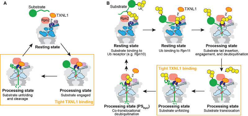
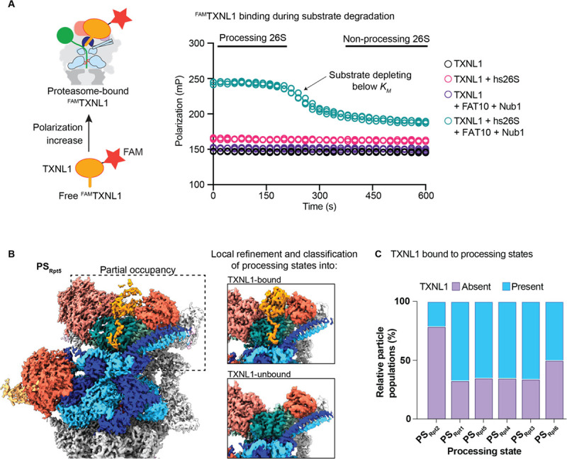
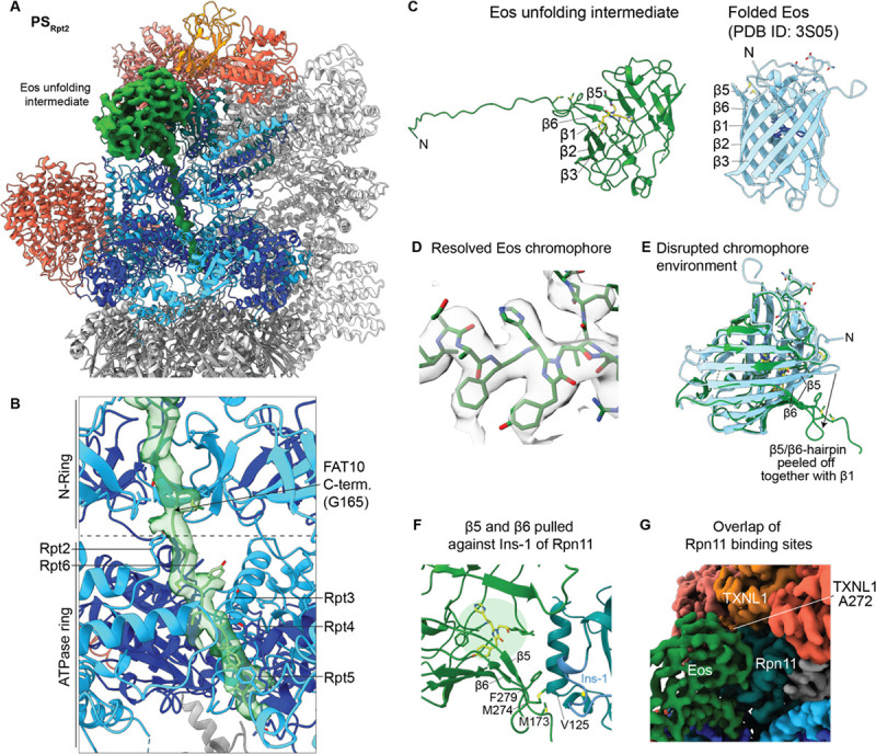
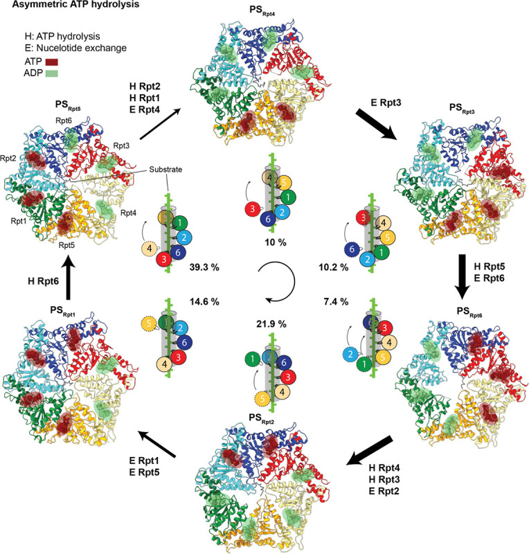
The 26S proteasome targets many cellular proteins for degradation during general homeostasis, protein quality control, and the regulation of vital processes. A broad range of proteasome-interacting cofactors thereby modulates these functions and aids in substrate degradation. Here, we solved several high-resolution structures of the redox active cofactor TXNL1 bound to the human 26S proteasome at saturating and sub-stoichiometric concentrations by time resolved cryo-EM. We identified distinct binding modes of TXNL1 that depend on the proteasome conformational and ATPase motor states. Together with biophysical and biochemical experiments, our structural studies reveal that the resting-state proteasome prior to substrate engagement with the ATPase motor binds TXNL1 with low affinity and in variable positions on top of the Rpn11 deubiquitinase. In contrast, the actively degrading proteasome shows additional interactions leading to high-affinity TXNL1 binding, whereby TXNL1's C-terminal tail covers the catalytic groove of the Rpn11 deubiquitinase and coordinates the active-site Zn. Furthermore, these cryo-EM structures of the degrading proteasome capture the ATPase hexamer in all registers of spiral-staircase arrangements and thus visualize the complete ATP-hydrolysis cycle of the AAA+ motor, indicating temporally asymmetric hydrolysis and conformational changes in bursts during mechanical substrate unfolding and translocation. Remarkably, we catch the proteasome in the act of unfolding the beta-barrel mEos3.2 substrate while the ATPase hexamer is in a particular spiral staircase register. Our findings challenge current models for protein translocation through hexameric AAA+ motors and reveal how the proteasome uses its distinct but broad range of conformational states to coordinate cofactor binding and substrate processing.

摘要

在一般的体内平衡、蛋白质质量控制和重要过程的调节过程中,26S蛋白酶体靶向许多细胞蛋白进行降解。因此,多种与蛋白酶体相互作用的辅因子调节这些功能并协助底物降解。在这里,我们通过时间分辨冷冻电镜解析了氧化还原活性辅因子TXNL1在饱和和亚化学计量浓度下与人26S蛋白酶体结合的几个高分辨率结构。我们确定了TXNL1不同的结合模式,这些模式取决于蛋白酶体的构象和ATP酶马达状态。结合生物物理和生化实验,我们的结构研究表明,在底物与ATP酶马达结合之前的静止状态蛋白酶体以低亲和力结合TXNL1,且结合位置在Rpn11去泛素酶顶部可变。相反,处于活跃降解状态的蛋白酶体显示出额外的相互作用,导致与TXNL1高亲和力结合,其中TXNL1的C末端尾巴覆盖Rpn11去泛素酶的催化凹槽并配位活性位点的锌。此外,这些降解状态蛋白酶体的冷冻电镜结构捕捉到了处于螺旋楼梯排列所有寄存器状态的ATP酶六聚体,从而可视化了AAA+马达完整的ATP水解循环,表明在机械性底物解折叠和转运过程中,ATP水解和构象变化在爆发时有时间上的不对称性。值得注意的是,当ATP酶六聚体处于特定的螺旋楼梯寄存器状态时,我们捕捉到蛋白酶体正在解折叠β桶状mEos3.2底物。我们的发现挑战了当前关于蛋白质通过六聚体AAA+马达转运的模型,并揭示了蛋白酶体如何利用其独特但广泛的构象状态来协调辅因子结合和底物加工。

https://cdn.ncbi.nlm.nih.gov/pmc/blobs/add5/11580985/235b59f7def8/nihpp-2024.11.08.622731v1-f0006.jpg
https://cdn.ncbi.nlm.nih.gov/pmc/blobs/add5/11580985/c58992cbc702/nihpp-2024.11.08.622731v1-f0007.jpg
https://cdn.ncbi.nlm.nih.gov/pmc/blobs/add5/11580985/8be71fe005cc/nihpp-2024.11.08.622731v1-f0008.jpg
https://cdn.ncbi.nlm.nih.gov/pmc/blobs/add5/11580985/b44e88b06bcb/nihpp-2024.11.08.622731v1-f0009.jpg
https://cdn.ncbi.nlm.nih.gov/pmc/blobs/add5/11580985/1ea02669429a/nihpp-2024.11.08.622731v1-f0010.jpg
https://cdn.ncbi.nlm.nih.gov/pmc/blobs/add5/11580985/87349a9bc95f/nihpp-2024.11.08.622731v1-f0011.jpg
https://cdn.ncbi.nlm.nih.gov/pmc/blobs/add5/11580985/99bce02a7142/nihpp-2024.11.08.622731v1-f0012.jpg
https://cdn.ncbi.nlm.nih.gov/pmc/blobs/add5/11580985/7e85babb8b46/nihpp-2024.11.08.622731v1-f0013.jpg
https://cdn.ncbi.nlm.nih.gov/pmc/blobs/add5/11580985/8b49224c0760/nihpp-2024.11.08.622731v1-f0014.jpg
https://cdn.ncbi.nlm.nih.gov/pmc/blobs/add5/11580985/a52807bd1e58/nihpp-2024.11.08.622731v1-f0015.jpg
https://cdn.ncbi.nlm.nih.gov/pmc/blobs/add5/11580985/abe5325e559e/nihpp-2024.11.08.622731v1-f0001.jpg
https://cdn.ncbi.nlm.nih.gov/pmc/blobs/add5/11580985/51417ac886ed/nihpp-2024.11.08.622731v1-f0002.jpg
https://cdn.ncbi.nlm.nih.gov/pmc/blobs/add5/11580985/7b23d68a6c5a/nihpp-2024.11.08.622731v1-f0003.jpg
https://cdn.ncbi.nlm.nih.gov/pmc/blobs/add5/11580985/87ff4fc87027/nihpp-2024.11.08.622731v1-f0004.jpg
https://cdn.ncbi.nlm.nih.gov/pmc/blobs/add5/11580985/6b8ba7101e24/nihpp-2024.11.08.622731v1-f0005.jpg
https://cdn.ncbi.nlm.nih.gov/pmc/blobs/add5/11580985/235b59f7def8/nihpp-2024.11.08.622731v1-f0006.jpg

相似文献

1
Structural landscape of AAA+ ATPase motor states in the substrate-degrading human 26S proteasome reveals conformation-specific binding of TXNL1.底物降解型人26S蛋白酶体中AAA+ATP酶运动状态的结构图谱揭示了TXNL1的构象特异性结合。
bioRxiv. 2024 Nov 9:2024.11.08.622731. doi: 10.1101/2024.11.08.622731.
2
Stairway to translocation: AAA+ motor structures reveal the mechanisms of ATP-dependent substrate translocation.易位的阶梯:AAA+ 马达结构揭示了 ATP 依赖的底物易位的机制。
Protein Sci. 2020 Feb;29(2):407-419. doi: 10.1002/pro.3743. Epub 2019 Oct 17.
3
The deubiquitinase Rpn11 functions as an allosteric ubiquitin sensor to promote substrate engagement by the 26S proteasome.去泛素化酶Rpn11作为一种变构泛素传感器,促进26S蛋白酶体与底物的结合。
Cell Rep. 2025 Jun 24;44(6):115736. doi: 10.1016/j.celrep.2025.115736. Epub 2025 May 22.
4
USP14-regulated allostery of the human proteasome by time-resolved cryo-EM.USP14 通过时间分辨冷冻电镜调节人蛋白酶体的变构。
Nature. 2022 May;605(7910):567-574. doi: 10.1038/s41586-022-04671-8. Epub 2022 Apr 27.
5
Structure, Dynamics and Function of the 26S Proteasome.26S 蛋白酶体的结构、动态与功能。
Subcell Biochem. 2021;96:1-151. doi: 10.1007/978-3-030-58971-4_1.
6
The deubiquitinase Rpn11 functions as an allosteric ubiquitin sensor to promote substrate engagement by the 26S proteasome.去泛素化酶Rpn11作为一种变构泛素传感器,促进26S蛋白酶体与底物的结合。
bioRxiv. 2024 Oct 24:2024.10.24.620116. doi: 10.1101/2024.10.24.620116.
7
An AAA Motor-Driven Mechanical Switch in Rpn11 Controls Deubiquitination at the 26S Proteasome.Rpn11 中的 AAA 马达驱动机械开关控制 26S 蛋白酶体的去泛素化作用。
Mol Cell. 2017 Sep 7;67(5):799-811.e8. doi: 10.1016/j.molcel.2017.07.023. Epub 2017 Aug 24.
8
Substrate-engaged 26 proteasome structures reveal mechanisms for ATP-hydrolysis-driven translocation.结合底物的 26 蛋白酶体结构揭示了 ATP 水解驱动转运的机制。
Science. 2018 Nov 30;362(6418). doi: 10.1126/science.aav0725. Epub 2018 Oct 11.
9
AAA+ ATPases in Protein Degradation: Structures, Functions and Mechanisms.AAA+ ATPases 在蛋白质降解中的作用:结构、功能与机制。
Biomolecules. 2020 Apr 18;10(4):629. doi: 10.3390/biom10040629.
10
An empirical energy landscape reveals mechanism of proteasome in polypeptide translocation.经验能景揭示蛋白酶体在多肽易位中的作用机制。
Elife. 2022 Jan 20;11:e71911. doi: 10.7554/eLife.71911.

本文引用的文献

1
The deubiquitinase Rpn11 functions as an allosteric ubiquitin sensor to promote substrate engagement by the 26S proteasome.去泛素化酶Rpn11作为一种变构泛素传感器,促进26S蛋白酶体与底物的结合。
Cell Rep. 2025 Jun 24;44(6):115736. doi: 10.1016/j.celrep.2025.115736. Epub 2025 May 22.
2
NUB1 traps unfolded FAT10 for ubiquitin-independent degradation by the 26S proteasome.NUB1捕获未折叠的FAT10,以便通过26S蛋白酶体进行非泛素依赖性降解。
Nat Struct Mol Biol. 2025 Apr 11. doi: 10.1038/s41594-025-01527-3.
3
Multi-pass, single-molecule nanopore reading of long protein strands.
多步、单分子纳米孔对长蛋白链的读取。
Nature. 2024 Sep;633(8030):662-669. doi: 10.1038/s41586-024-07935-7. Epub 2024 Sep 11.
4
The midnolin-proteasome pathway catches proteins for ubiquitination-independent degradation.中间层蛋白酶体途径捕获蛋白质进行非依赖泛素化的降解。
Science. 2023 Aug 25;381(6660):eadh5021. doi: 10.1126/science.adh5021.
5
Ubiquitin modulates 26 proteasome conformational dynamics and promotes substrate degradation.泛素调节 26S 蛋白酶体构象动力学并促进底物降解。
Sci Adv. 2022 Dec 23;8(51):eadd9520. doi: 10.1126/sciadv.add9520.
6
USP14-regulated allostery of the human proteasome by time-resolved cryo-EM.USP14 通过时间分辨冷冻电镜调节人蛋白酶体的变构。
Nature. 2022 May;605(7910):567-574. doi: 10.1038/s41586-022-04671-8. Epub 2022 Apr 27.
7
DeepEMhancer: a deep learning solution for cryo-EM volume post-processing.DeepEMhancer:一种用于冷冻电镜体积后处理的深度学习解决方案。
Commun Biol. 2021 Jul 15;4(1):874. doi: 10.1038/s42003-021-02399-1.
8
Highly accurate protein structure prediction with AlphaFold.利用 AlphaFold 进行高精度蛋白质结构预测。
Nature. 2021 Aug;596(7873):583-589. doi: 10.1038/s41586-021-03819-2. Epub 2021 Jul 15.
9
3D variability analysis: Resolving continuous flexibility and discrete heterogeneity from single particle cryo-EM.3D 变异性分析:从单颗粒冷冻电镜中解析连续的柔韧性和离散的异质性。
J Struct Biol. 2021 Jun;213(2):107702. doi: 10.1016/j.jsb.2021.107702. Epub 2021 Feb 11.
10
Macromolecular structure determination using X-rays, neutrons and electrons: recent developments in Phenix.利用 X 射线、中子和电子进行高分子结构测定: Phenix 的最新进展。
Acta Crystallogr D Struct Biol. 2019 Oct 1;75(Pt 10):861-877. doi: 10.1107/S2059798319011471. Epub 2019 Oct 2.